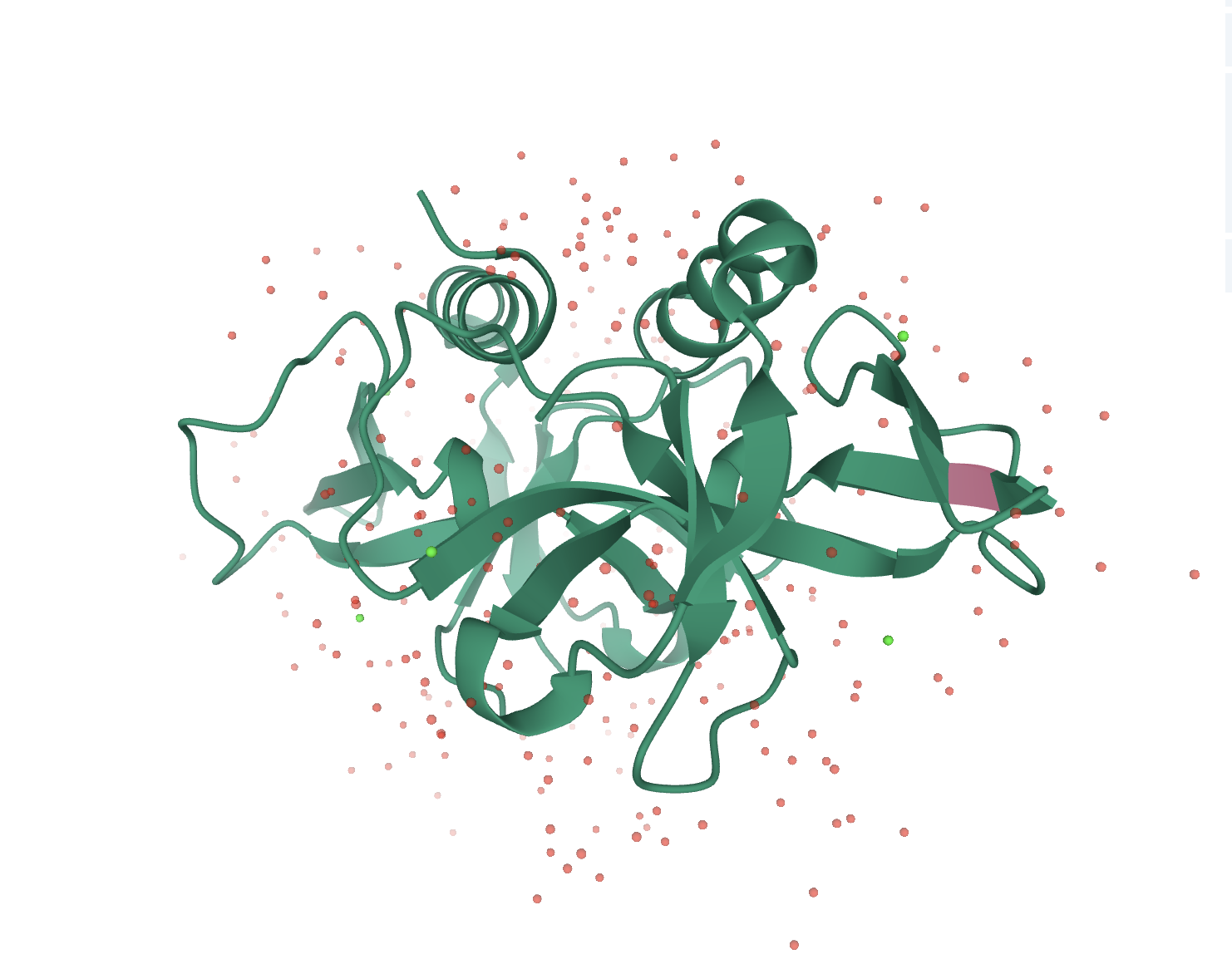
Part:BBa_K3512001
ccdB Toxin
The CcdB toxin is part of the CcdA-CcdB system. The target of CcdB is the GyrA subunit of DNA gyrase, an essential type II topoisomerase in Escherichia coli. Gyrase alters DNA topology by effecting a transient double-strand break in the DNA backbone, passing the double helix through the gate and resealing the gaps. The CcdB toxin acts by trapping DNA gyrase in a cleaved complex with the gyrase A subunit covalently closed to the cleaved DNA, causing DNA breakage and cell death in a way closely related to quinolones antibiotics.
In absence of the antitoxin CcdA, the CcdB toxin traps DNA-gyrase cleavable complexes, inducing breaks into DNA and cell death.
3D View
Contribution from Bioplus-China 2022
Introduction
Our team inquired about a literature, sorted out the results, and supplemented BBa_ K3512001 with relevant information. The origin and the evolution of toxin-antitoxin systems (TA) remain to be uncovered. TA are abundant in bacterial chromosomes and are thought to be part of the flexible genome that originates from horizontal gene transfer (HGT). To gain insight into TA evolution, They analysed the distribution of the chromosomally-encoded ccdO157 system in 395 natural isolates of E. coli. It was discovered in the E. coli O157:H7 strain in which it constitutes a genomic islet between 2 core genes (folA and apaH). This literature revealed that Molecular evolution analysis showed that ccdBO157 is under neutral evolution, suggesting that this system is devoid of any biological role in the E. coli species.
Method
Toxicity and antitoxicity plate assays To test the toxicity of the cloned CcdBO157 and CcdBF variants, the corresponding pBAD33-ccdB constructs were transformed in MG1655. The resulting transformants were plated on LB plates containing chloramphenicol with or without arabinose (1%). The CcdB variants were considered to be functional (toxic) when transformants were able to grow only in the absence of arabinose. To test the ability of the cloned CcdAO157 and CcdAF variants to counteract the toxicity of CcdBO157 and CcdBF, respectively, the corresponding pKK-ccdA constructs were transformed in MG1655 expressing the reference ccdBO157 orccdBF genes from the pBAD33 vector. The resulting transformants were plated on LB plates containing chloramphenicol and ampicillin with arabinose (1%). Basal expression of ccdA from the pTac promoter of pKK223-3 in MG1655 is sufficient to test the antitoxicity phenotype. The CcdA variants were considered to be functional when the toxicity of CcdBO157 or CcdBF protein was counteracted, i.e., when strains coexpressing a ccdA variant with the ccdBO157 or ccdBF reference genes were able to grow in the presence of arabinose while strains expressing only the ccdBO157 or ccdBF reference gene were not.
Results
Variants of CcdB In this paper, they found that the CcdB toxin proteins were much more diversified than the antitoxins. Among the 47 serogroups, 14 classes of alleles could be identified. Note that, as mentioned earlier, only one isolate for each serogroup was sequenced and tested (47 isolates). One class was composed of 4 isolates presenting sequence identical to the ccdBO157 gene of the O157:H7 EDL933 reference strain. The 2 most prevalent classes represented 13/47 and 8/47 isolates, and the corresponding alleles harboured either two variations (S10G and V28E) or 1 variation (S44I), respectively. The toxic activity of at least one representative CcdB protein of each class was tested using the toxicity plate assay (see materials and methods) and was comparable to that of the CcdBO157 protein. Four classes, together representing 7 isolates (7/47), contained from one to up to 5 variations (V28E; RH7-V28E; S10G-I26V-V28E; RH7-S10G-I26V-V28E- I93L). At least one representative of each class was assayed for toxicity using the toxicity plate assay. Interestingly, expression of these variants led to cell killing, showing that the variations did not affect the toxic activity of the CcdB proteins. On the 7 classes remaining, 1 is composed of 2 isolates (representing serogroups O138 and O153) containing 4 variations in their ccdB gene (S10G-V28E-S44I-P54T). The CcdBO138 protein (from serogroup O138) was assayed for toxicity and surprisingly shown not to affect viability upon ectopic expression (Figure 1). The comparison of the variations among the different classes suggests that the P54T mutation is responsible for abolishing the toxic activity of the CcdBO138 variant. The 6 last classes, representing 13/47 isolates, were composed of truncated proteins. Deletions of the carboxy-terminal region were caused by amber mutations at various locations (E41, R42, E63, C84). Interestingly, all these truncated proteins contained several point variations that were also found in the full-length variants that were still toxic (S10G, I26V, V28E, S44I) or not (R7H, P54T). Two of these classes (7/47 isolates) contained 1 extra variation (K62S), while another class (1/47 isolates) contained 2 more variations (R11S and R32S). One representative of 4 out of these 6 classes was tested and was shown to be non toxic using the plate toxicity assay (data not shown). Figure 1 shows that ectopic overexpression of CcdBO51 in the liquid toxicity assay did not lead to the loss of viability. These truncated variants of CcdBO157 are thus inactive and it is likely to be the case for CcdBO7 and CcdBO102 since the active site of the CcdBO157 is located at the caboxy-terminus of the protein (BAHASSI et al. 1995; WILBAUX et al. 2007). The corresponding antitoxins were functional (Table 1).


Contribution From XMU-China 2023
The ccdB gene is located on the Escherichia coli F-factor plasmid and it is a part of the toxin-antitoxin system encoded by theccd operon. This part (BBa_K3512001) was first registered in 2020 (Team: http://BITSPilani-Goa India/Parts - 2020.igem.org) and used as a toxin in biosafety applications. For the contribution of this part, the survival assay (1) was implemented to characterize the effect of the toxin, and the results were added to the page of the corresponding part.
Construction of gene circuit
We used pBAD/araC (BBa_I0500), RBS (BBa_B0034), ccdB (BBa_K3512001) to construct the composite part (BBa_K4907140), which were assembled on pSB4K5 backbone by standard assembly (Fig. 1).

This constructed circuit was transformed into E. coli DH10β, followed by positive transformant selection using kanamycin and confirmation through colony PCR (Fig. 2) and sequencing.

Characterization of killing effect
We used promoter BBa_I0500 to regulate the expression of CcdB. After induction by L-arabinose, OD600 values were measured every two hours. The bacteria that had no CcdB expressed grew rapidly, while the one expressing CcdB showed a significant growth defect, as the optical density (at 600 nm) increased very slightly (Fig. 3a).

At each time, the spot assay was also performed, then the cell viability was measured by CFU count (Fig. 3b). Consistent with the trend of OD600 value against time, only the absence of CcdB allowed the host cells to survive. All these results indicated that CcdB was toxic enough to the engineered bacteria so that this toxin could be applied to the cases when the suicide of genetically engineered microorganisms (GEMs) were strongly needed.
The neutralisation of CcdA to CcdB
ccdA is another gene in the ccd operon that encodes the anti-toxic protein CcdA, which protects cells from the toxic effect of CcdB via neutralisation. DB3.1 Super Competent Cell is a kind of E. coli which is capable of efficiently expanding the plasmid carrying bacterial toxic gene ccdB cause the ccdA is located on its genome. To validate the neutralization of ccdA to ccdB, we performed a series of dual-plasmid transformations to different E. coli strains as shown in Table. 1. The correct dual-plasmid system was confirmed by chloramphenicol and kanamycin.
| Experiment | Dual-plasmid system | Strain | Result | Colonies |
|---|---|---|---|---|
| Verfication of ccdA in plasmid | BBa_K4907139_pSB4K5 BBa_K4907138_pSB1C3 |
in DH10β |
 |
✔ |
| BBa_K4907139_pSB4K5 BBa_I13453_pSB1C3 |
 |
✖️ | ||
| Verfication of ccdA in genome (ccdB is controlled by pRha) | BBa_K4907131_pSB4K5 BBa_I13453_pSB1C3 |
in DB3.1 |
 |
✔ |
| in DH10β |
 |
✖️ | ||
| Verfication of ccdA in genome (ccdB is controlled by pBAD) | BBa_K4907139_pSB4K5 BBa_I13453_pSB1C3 |
in DB3.1 |
 |
✔ |
| in DH10β |
 |
✖️ |
The results showed that E. coli DB3.1 transformed with toxin genes andE. coli DH10β transformed with pBAD (BBa_K206001) promoter or pRha (BBa_K914003) promoter grew well. E. coli DH10β transformed with both toxins and antitoxins also exhibited good growth. However, E. coli DH10β with toxins did not grow. This confirmed the killing effect of the toxin CcdB again and the neutralization of antitoxin CcdA. Besides, the E. coli DB3.1 transformed with toxin controlled by pBAD promoter or pRha promoter and without antitoxin, both grew better compared with E. coli DH10β. From these results, we can draw the conclusion that whether the ccdA is in plasmid or genome can play the role of neutralization to ccdB.
Reference
Mine, N., Guglielmini, J., Wilbaux, M. and Van Melderen, L., 2009. The Decay of the Chromosomally Encoded ccdO157 Toxin–Antitoxin System in the Escherichia coli Species. Genetics, 181(4), pp.1557-1566.
C. T. Chan, J. W. Lee, D. E. Cameron, C. J. Bashor, J. J. Collins, 'Deadman' and 'Passcode' microbial kill switches for bacterial containment. Nat Chem Biol. 12, 82-86 (2016).
Sequence and Features
- 10COMPATIBLE WITH RFC[10]
- 12COMPATIBLE WITH RFC[12]
- 21COMPATIBLE WITH RFC[21]
- 23COMPATIBLE WITH RFC[23]
- 25COMPATIBLE WITH RFC[25]
- 1000INCOMPATIBLE WITH RFC[1000]Illegal BsaI site found at 216
//biosafety/kill_switch
//function/celldeath
| dissociation_constant | High affinity = 3.57*10^-3 nM |
| protein | Dissociation complex = 1.36*10^-9 nM |

