Part:BBa_K1172909
Biosafety-System araCtive (AraC)
The Biosafety-System araCtive BBa_K1172909 is an improvement of the BioBrick BBa_K914014 by replacing:
- the first promoter arabinose PBAD by the rhamnose promoter PRha to obtain a lower basal transcription.
- intgration of the alanine racemase BBa_K1172901(alr) for gaining higher plasmid stability and for taking advantage of the double-kill switch.
- the repressor LacI to AraC and the lac promoter to the arabinose promoter PBAD for a higher repression and lower basal transcription of the second part.
For more details about the seperate genes in this Biosafety-System and their function, click [http://2013.igem.org/Team:Bielefeld-Germany/Biosafety/Biosafety_System_S here]
For more details about the function of the Biosafety-System in general, click [http://2013.igem.org/Team:Bielefeld-Germany/Biosafety/Biosafety_System here]
Sequence and Features
- 10COMPATIBLE WITH RFC[10]
- 12INCOMPATIBLE WITH RFC[12]Illegal NheI site found at 1374
Illegal NheI site found at 2393 - 21INCOMPATIBLE WITH RFC[21]Illegal BglII site found at 1298
Illegal BamHI site found at 2000
Illegal BamHI site found at 2333 - 23COMPATIBLE WITH RFC[23]
- 25INCOMPATIBLE WITH RFC[25]Illegal AgeI site found at 1416
Illegal AgeI site found at 1716 - 1000INCOMPATIBLE WITH RFC[1000]Illegal BsaI.rc site found at 1173
Illegal BsaI.rc site found at 3068
Biosafety-System araCtive
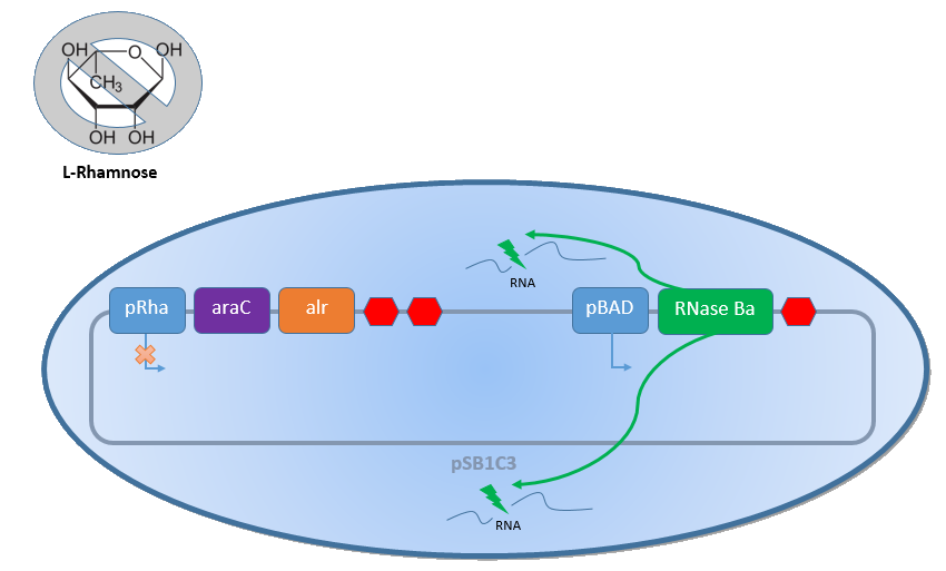
Combining the [http://2013.igem.org/Team:Bielefeld-Germany/Biosafety/Biosafety_System_S genes] described above with the Biosafety-Strain K-12 ∆alr ∆dadX results in a powerful device, allowing us to control the bacterial cell division. The control of the bacterial growth is possible either active or passive. Active by inducing the PBAD promoter with L-arabinose and passive by the induction of L-rhamnose. The passive control makes it possible to control the bacterial cell division in a defined closed environment, like the MFC, by continuously adding L-rhamnose to the medium. As shown in the Figure 1 below, this leads to an expression of the essential alanine racemase (alr) and the AraC regulator, so that the expression of the RNase Ba is repressed.
In the event that bacteria exit the defined environment of the MFC or L-rhamnose is not added to the medium any more, both the expression of the alanine racemase (Alr) and the AraC regulatordecrease, so that the expression of the toxic RNase Ba (Barnase) begins. The cleavage of the intracellular RNA by the Barnase and the lack of synthesized D-alanine, caused by the repressed alanine racemase inhibit the cell division and makes sure that the bacteria can only grow in the defined environment or the device of choice respectively.

Results
Characterization of the arabinose promoter PBAD
First of all, the arabinose promoter PBAD was characterized to get a first impression of its basal transcription rate. Therefore the bacterial growth was investigated under the pressure of the unrepressed PBAD promoter on different carbon source using M9 minimal medium with either glucose or glycerol. The transcription rate was determined by fluorescence measurement of GFP BBa_E0040 transcribed via the PBAD promoter using the BioBrick BBa_I13541.
As shown in Figure 3 below, the bacteria grew better on glucose than on glycerol. This is caused by the fact that glucose is the better energy source of this two, because glycerol enters glycolysis at a later step and therefore delivers less energy (in addition to the additional ATP consumption to drive glycerol uptake). For the investigation of the basal transcription the fluorescence measurements, shown in Figure 4, is more interesting.
Both the wild type E. coli K-12 and the strain E. coli K-12 ∆alr ∆dadX ∆araC show about the same fluorescence on glucose (red and orange curve) but differ on glycerol. This can be explained by the fact that growth on glucose does not cause an additional induction of the arabinose promoter PBAD, while glycerol does. In the presence of glucose, the intracellular concentration of cAMP is low. The absence of cAMP results in inactive CAP (carbon utilization activator protein) and thus no induction of inefficient catabolisms like the catabolism of arabinose. Therefore, glucose is catabolized by the bacteria first, resulting in an optimal growth. In the absence of glucose, the cAMP level increases which enhances via CAP-cAMP the transcription of most operons encoding enzymes for the catabolism of alternative carbon sources. Therefore, the expression of GFP under the control of the PBAD promoter increases on glycerol. Another difference can be seen between the wild type (black curve) and the strain K-12 ∆alr ∆dadX ∆araC (blue curve).
The Biosafety-Strain shows lower expression then the wild type, but has about the same growth rate, according to Figure 11. The reason for this characteristic is caused by the deleted AraC protein in the Biosafety-Strain. Because the AraC protein functions not only as are repressor but also as an activator the transcription rate decreases in the Biosafety-Strain.


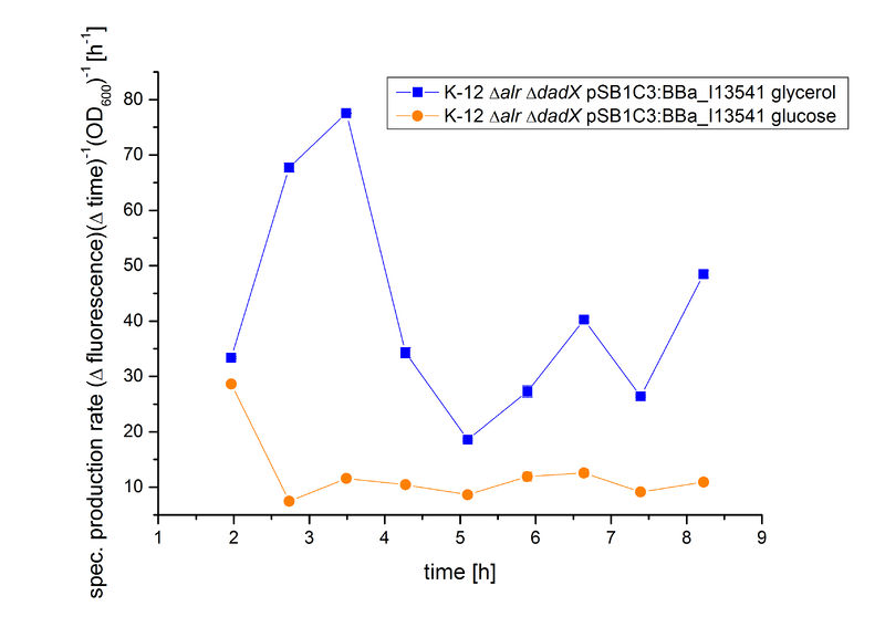
The effect that glucose represses the transcription of the PBAD promoter becomes more obvious by calculating of the specific production rates of GFP, as shown in Figure 5. The specific production rates were calculated via equation (1) :
With the specific production rate of GFP, it can be demonstrated that GFP synthesis rates differ extremely between the cultivation on glucose and glycerol. The specific production rate is constantly very low when using glucose as carbon source, but shows a higher more unstable level in the cultivation with glycerol.
Because the specific production rate was calculated between every single measurement point, the curve in Figure 5 is not smoothed and so the fluctuations have to be ignored, as they do not stand for are real fluctuations in the expression of GFP. They are caused by measurement variations in the growth curve and the fluorescence curve. And as neither of this curves are ideal, the fluctuations are the result. Nevertheless this graph shows clearly the difference between the two carbon sources.
Characterization of the Biosafety-System araCtive
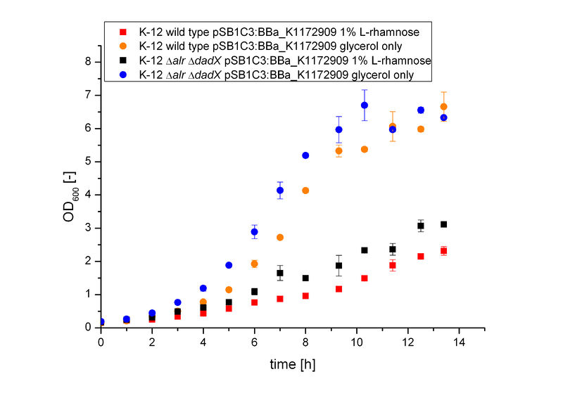
The Biosafety-System araCtive was characterized on M9 minimal medium using glycerol as carbon source. As for the characterization of the pure arabinose promoter PBAD above, the bacterial growth and the fluorescence of GFP BBa_E0040 was measured. Therefore, the wild type and the Biosafety-Strain E. coli K-12 ∆alr ∆dadX, both containing the Biosafety-Plasmid BBa_K1172909, were cultivated once with the induction of 1% L-rhamnose and once only on glycerol.
It becomes obvious (Figure 6) that the bacteria, induced with 1 % L-rhamnose (red and black curve) grow significantly slower than on pure glycerol (orange and blue curve). This is attributed to the high metabolic burden encountered by the induced bacteria. The expression of the repressor AraC and the alanine racemase (Alr) simultaneously causes a high strain on the cells, so that they grow slower than the uninduced cells, which express only GFP.
Comparing the bacterial growth with the fluorescence in Figure 7, it can be seen that the fluorescence seems to follow the same trend than the bacterial growth. The uninduced cells show approximately an exponential rise of fluorescence, while in comparision the fluorescence of the induced bacteria increases only slowly.


From the data presented above it can not be determined if the expression of the repressor AraC does affect the transcription of GFP or not. The slower growth of the bacteria is a first indication that the repressor AraC and the alanine racemase (Alr) are highly expressed, but the growth of the bacteria shows nearly the same kinetics as the fluorescence. So it could be possible that the repressor does not affect the expression level of GFP under the control of the arabinose promoter PBAD. This becomes more clear by the calculation of the specific production rate of GFP by equation (1) . As shown in Figure 8 below the specific production rate differs clearly between the uninduced Biosafety-System and the Biosafety-System induced by 1% L-rhamnose. The production of GFP in the presence of L-rhamnose (red curve) is always lower than in its absence (orange curve), so that the expression of GFP is repressed in the presence of L-rhamnose.
Because the specific production rate of GFP was calculated between every single measurement point, the curve in Figure 8 is not smoothed and so the fluctuations have to be ignored, as they do not stand for are real fluctuations in the expression of GFP. They are caused by measurement variations in the growth curve and the fluorescence curve. But there is a clear tendency that the production of GFP is significantly lower when the bacteria are induced with 1% L-rhamnose. So the Biosafety-System araCtive works.
Conclusion of the Results
For the conclusion of the result, visit our [http://2013.igem.org/Team:Bielefeld-Germany/Biosafety/Biosafety_System_S#Conclusions wiki].
References
- Baldoma L and Aguilar J (1988) Metabolism of L-Fucose and L-Rhamnose in Escherichia coli: Aerobic-Anaerobic Regulation of L-Lactaldehyde Dissimilation [http://www.ncbi.nlm.nih.gov/pmc/articles/PMC210658/pdf/jbacter00179-0434.pdf|Journal of Bacteriology 170: 416 - 421.].
- Carafa, Yves d'Aubenton Brody, Edward and Claude (1990) Thermest Prediction of Rho-independent Escherichia coli Transcription Terminators - A Statistical Analysis of their RNA Stem-Loop Structures [http://ac.els-cdn.com/S0022283699800059/1-s2.0-S0022283699800059-main.pdf?_tid=ede07e2a-2a92-11e3-b889-00000aab0f6c&acdnat=1380629809_2d1a59e395fc69c8608ab8b5aea842f7|Journal of molecular biology 216: 835 - 858].
- Cass, Laura G. and Wilcoy, Gary (1988) Novel Activation of araC Expression and a DNA Site Required for araC Autoregulation in Escherichia coli B/r [http://www.ncbi.nlm.nih.gov/pmc/articles/PMC211425/pdf/jbacter00187-0394.pdf|Journal of Bacteriology 170: 4174 - 4180].
- Hamilton, Eileen P. and Lee, Nancy (1988) Three binding sites for AraC protein are required for autoregulation of araC in Escherichia coli [http://www.ncbi.nlm.nih.gov/pmc/articles/PMC279856/pdf/pnas00258-0029.pdf|Proc Natl Acad Sci U S A 85: 1749 - 53].
- Mossakowska, Danuta E. Nyberg, Kerstin and Fersht, Alan R. (1989) Kinetic Characterization of the Recombinant Ribonuclease from Bacillus amyloliquefaciens (Barnase) and Investigation of Key Residues in Catalysis by Site-Directed Mutagenesis [http://pubs.acs.org/doi/pdf/10.1021/bi00435a033|Biochemistry 28: 3843 - 3850.].
- Paddon, C. J. Vasantha, N. and Hartley, R. W. (1989) Translation and Processing of Bacillus amyloliquefaciens Extracellular Rnase [http://www.ncbi.nlm.nih.gov/pmc/articles/PMC209718/pdf/jbacter00168-0575.pdf|Journal of Bacteriology 171: 1185 - 1187.].
- Schleif, Robert (2010) AraC protein, regulation of the L-arabinose operon in Escherichia coli, and the light switch mechanism of AraC action [http://gene.bio.jhu.edu/Ourspdf/127.pdf|FEMS microbial reviews 34: 779 - 796.].
- Voss, Carsten Lindau, Dennis and Flaschel, Erwin (2006) Production of Recombinant RNase Ba and Its Application in Downstream Processing of Plasmid DNA for Pharmaceutical Use [http://onlinelibrary.wiley.com/doi/10.1021/bp050417e/pdf|Biotechnology Progress 22: 737 - 744.].
- Walsh, Christopher (1989) Enzymes in the D-alanine branch of bacterial cell wall peptidoglycan assembly. [http://www.jbc.org/content/264/5/2393.long|Journal of biological chemistry 264: 2393 - 2396.]
- Wickstrum, J.R., Santangelo, T.J., and Egan, S.M. (2005) Cyclic AMP receptor protein and RhaR synergistically activate transcription from the L-rhamnose-responsive rhaSR promoter in Escherichia coli. [http://www.ncbi.nlm.nih.gov/pmc/articles/PMC1251584/?report=reader|Journal of Bacteriology 187: 6708 – 6719.].
Biosafety Double Kill Switch function:
we conducted experiments to proof that the deprivation of L-Rhamnose activates the proposed Double Kill Switch system and induce cell death. E. coli with Kill Switch genes and without kill switch genes are nurtured in LB liquid culture respectively for 24 hours. From the beginning of the culturing, L-Rhamnose is added to both cultures. The culturing began at when both groups’ OD 600 equals 0.1. OD 600 is measured to indicate the concentration of bacteria body within the culture. At the 9th hour of culturing, L-Rhamnose is no longer added, and Rhamnase is added. The concentrations of control and test bacteria are compared. The result is represented in the graph:
Added by RDFZ-CHINA
The result shows that the bacteria with kill switch, after a lagging time for around 3 hours after rhamnose is no longer added and rhamnase is added, began to significantly decrease in concentration, whereas the control group that carry no kill switch genes were unaffected by the deprivation of rhamnose and continue to increase in concentration. The comparison of the bacteria’s performance successfully demonstrates that the kill switch effectively kills the bacteria when L-rhamnose is not present in the environment.
| None |


