Part:BBa_K3182100
Contents
Sequence and Features
- 10COMPATIBLE WITH RFC[10]
- 12INCOMPATIBLE WITH RFC[12]Illegal NheI site found at 592
Illegal NheI site found at 615 - 21INCOMPATIBLE WITH RFC[21]Illegal BamHI site found at 580
- 23COMPATIBLE WITH RFC[23]
- 25COMPATIBLE WITH RFC[25]
- 1000COMPATIBLE WITH RFC[1000]
Introduction
pT7-CBDcipA-pCons-AsPink

This part consists of a carbohydrate binding domain (CBD) from Clostridium thermocellum (C. thermocellum) cellulose scaffolding protein (CipA). This binding domain is a central part of Clostridium thermocellum's cellusome and has a strong affinity for cellulose. The CBD was fused to another protein using a flexible GS-linker (-GGGGSGGGGS-)in order to attach this complex to a polysaccaride material. A thrombin cleavage site (-LVPRGS-) was added to the end of the linker and its breakage will leave a glycine and serine attached to the N-terminal of the fusion protein. The main mechanism of iGEM19 Linköping's project can be seen in Figure 1.
Protease site and use
The thrombin site was added to enable the ability to release the fusion protein down into skin wounds. Thanks to our integrated human practice we learned that infections span much deeper into wounds that we thought. Simply attaching the CBD-fusion protein to a carbohydrate material would not enable the fusion protein to reach far into the wound. The thrombin site was also chosen because of thrombin's endogenous existence in humans.
Assembly compabilities
An internal BamHI recognition sequence (RS) has been added to enable interchangeable fusion proteins to the CBD. BamHI was chosen because its RS codes for glycine and serine, fitting it to the end of the thrombin site. It is also a cost-effective enzyme and is unaffected by methylated DNA. BamHI is a part of the RFC21 standard.
CBDcipA crystal structure
Important molecular faces
CBDcipA is composed of a nine-stranded beta sandwich with a jelly roll topology and binds a calcium ion, which can be seen in Figure 2. It further contains conserved residues exposed on the surface which map into two clear surfaces on each side of the molecule. One of the faces mainly contains planar strips of aromatic and polar residues which may be the carbohydrate binding part. Further aspects are unknown and unique to this CBD such as the other conserved residues which are contained in a groove.
Carbohydrate binding domain specificity
Since the CBD is from the cellusome of C. thermocellum some research labeled it a cellulose binding domain. However, iGEM19 Linköping noticed that this domain could also bind to different sources of polysaccaride materials. This serves as a domain for iGEM19 Linköpings modular bandage, where the polysaccaride material can be exchanged for other/similar materials and not exclusively cellulose.
The choice of carbohydrate binding domain
iGEM Linköping 2019 chose CBDcipA due to the fact that many other iGEM teams had explored the possibilities of this domain. Our basic design was influenced by [http://2014.igem.org/Team:Imperial iGEM14 Imperial], [http://2015.igem.org/Team:edinburgh iGEM15 Edinburgh] and [http://2018.igem.org/Team:ecuador iGEM18 Ecuador]. Purification and where to place the fusion protein (N- or C-terminal) was determined by studying the former projects. CBDcipA also originates from a thermophilic bacteria which further increases the domain's applications.
Expression system
The part has a very strong expression with a T7-RNA-polymerase promotor (BBa_I719005), seen in Figure 3, as well as a 5'-UTR (BBa_K1758100) region which has been shown to further increase expression in Escherichia coli (E. coli) (BBa_K1758106), ([http://www.ncbi.nlm.nih.gov/pubmed/2676996 Olins et al. 1989]), ([http://www.ncbi.nlm.nih.gov/pubmed/23927491 Takahashi et al. 2013]).

Theoretical usage of this part
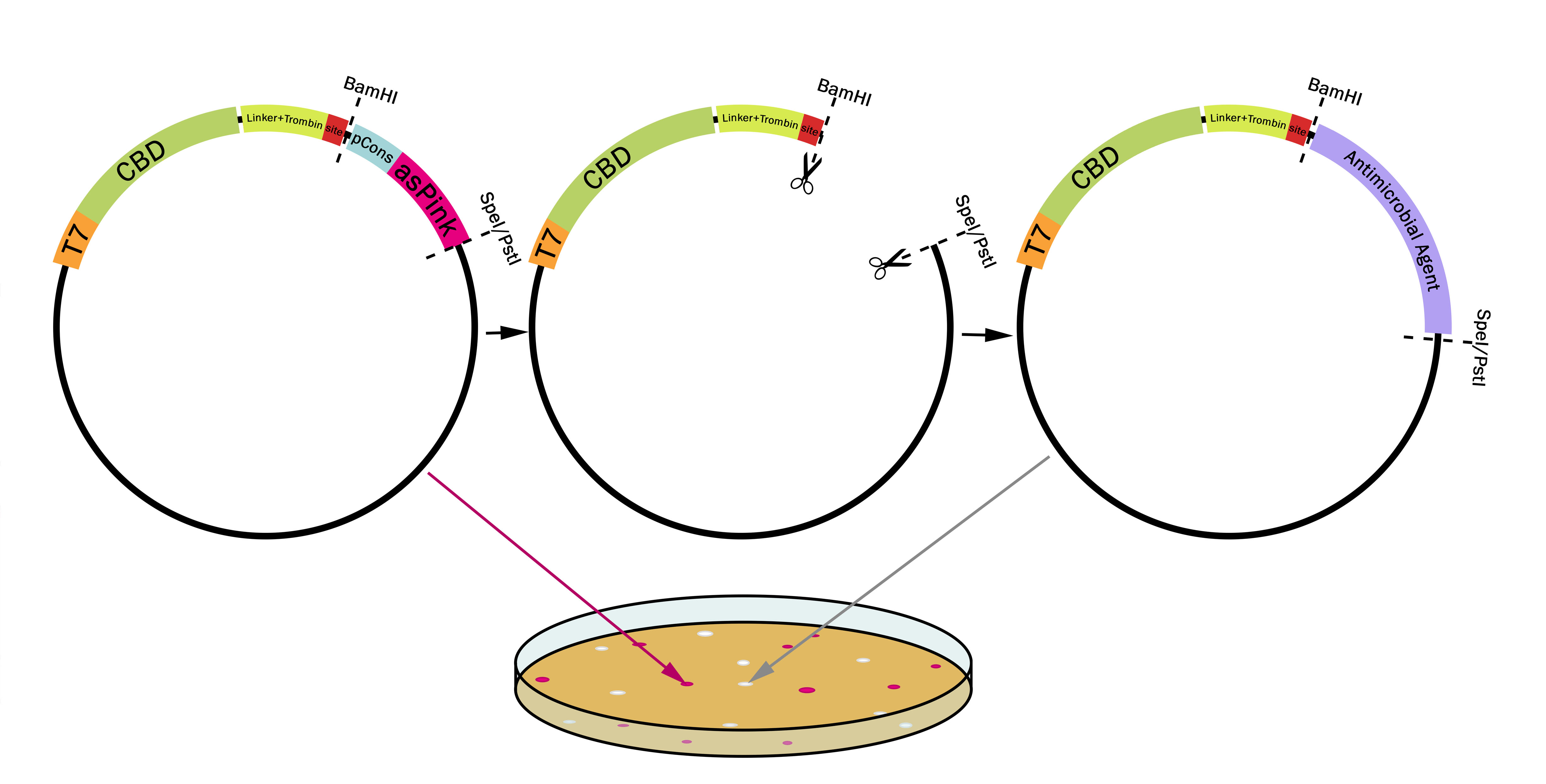
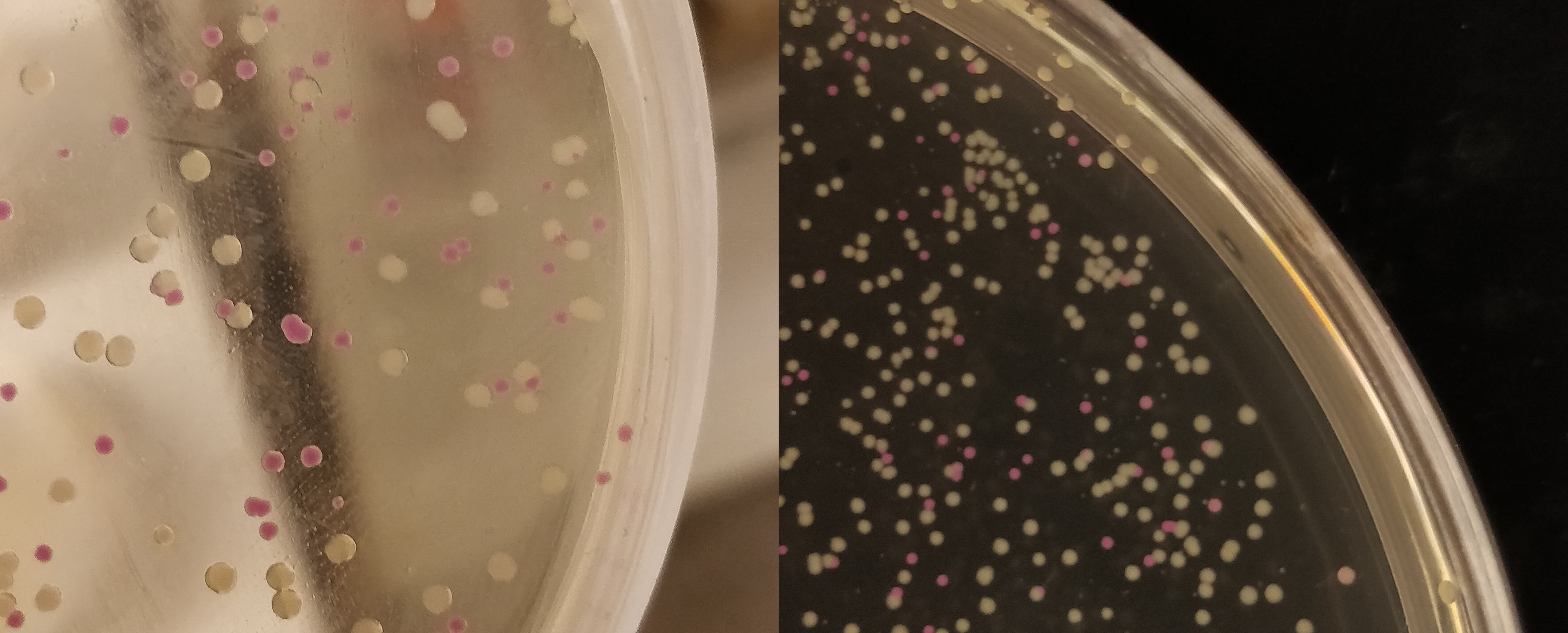
This part utilizes a pCons-AsPink dropout enabling colour-screening for positive colonies. Using BamHI and PstI or SpeI on both this part assembled in pSB1C3 (or vector of choice) and the insert of choice will yield a fusion protein between CBDcipA and the insert. The fusion protein can later be cleaved with thrombin to yield two separate proteins. The C-terminal fusion will have one glycine and one serine added to the N-terminal of the protein.

Examples of biobricks assembled with this method
This method was used to assemble: BBa_K3182103, BBa_K3182104, BBa_K3182105, BBa_K3182106, BBa_K3182107, BBa_K3182108 into pSB1C3. However, as we quickly noticed, pSB1C3 was not able to replicate in Vibrio natriegens (Vmax). After countless failed transformation attempts, all the above parts were also assembled into a modified pUC19 vector using the pink-white screening method. The pUC19 variants were transformed without any problems into V. natriegens (Vmax).
Usage and Biology
Design any gene which you want to be fused to the CBDcipA with a BamHI recognition sequence in the 5'-end. The biobrick suffix can be used in the 3'-end. Cut the vector and insert with BamHI and PstI (SpeI also works), remove enzymes and mix, no need for gel purification. Using very high molar ratios might not yield any pink colonies at all, a molar ratio insert to vector of 7-20:1 will yield some pink colonies. Transform the host (BL21 (DE3) for quickest results) and incubate at 37 degrees Celsius overnight, if the color is weak or can not be seen, incubate in 24-37 degrees Celsius for an additional 16-24 hours.

Colonies from the pink-white screening were later sent for sequencing, where all white colonies came back as correctly ligated plasmids. The only colony-screened assembly attemp can be seen in Figure X2, where all colonies screened showed the correct fragments, as well as all non-pink colonies showing green fluorescence. After the results from X2 had been sequenced, no more colony screening had to be done. All white colonies sent to sequencing contained the correctly assembled fusion proteins.
| None |


