Coding
mRFP1

Part:BBa_E1010

Designed by: Drew Endy   Group: Antiquity   (2004-07-28)
Revision as of 04:56, 30 October 2017 by Herlohuang (Talk | contribs)

**highly** engineered mutant of red fluorescent protein from Discosoma striata (coral)

monomeric RFP: Red Fluorescent Protein. Excitation peak: 584 nm Emission peak: 607 nm

Usage and Biology

Robert E. Campbell started with Discosoma RFP (DsRed) and evolved a faster folding, monomeric variant. See paper listed in source. Codon optimized for expression in bacteria (?? DE)

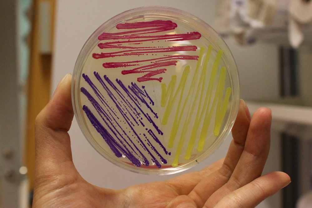
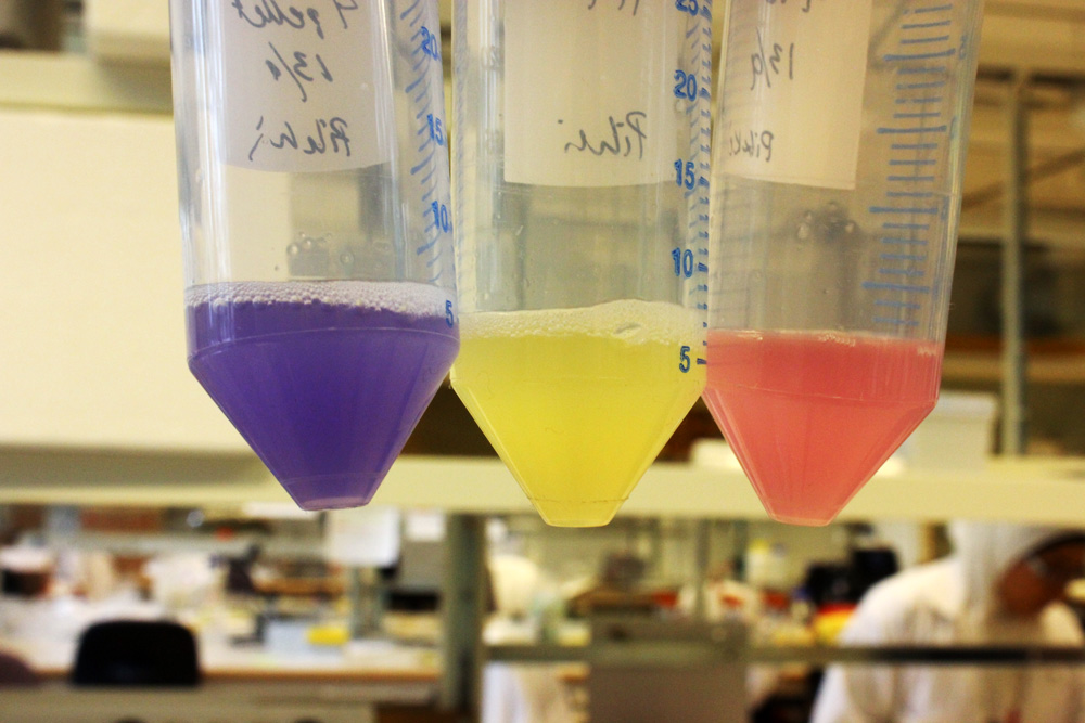
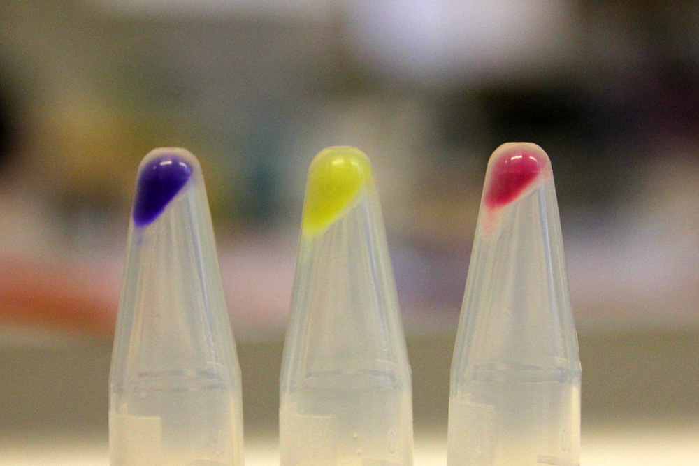
AmilCP amilGFP RFP.jpg On cultures BYR small.jpg Pellets BYR.jpg

iGEM11_Uppsala-Sweden: Expression of chromoproteins. The images above show E coli constitutively expressing amilCP BBa_K592009 (blue), amilGFP BBa_K592010 (yellow) and RFP BBa_E1010 (red).

Peking iGEM 2016 has fused this part with triple spytag. The fused protein is participate in Peking’s polymer network. By adding this protein, the whole polymer network become visible in most conditions. If you want to learn more about Peking’s polymer network and the role of mRFP in this network, please click here https://parts.igem.org/Part:BBa_K1989004".


Contribution

Group: Hong Kong-CUHK iGEM 2017
Author: Yuet Ching Lin
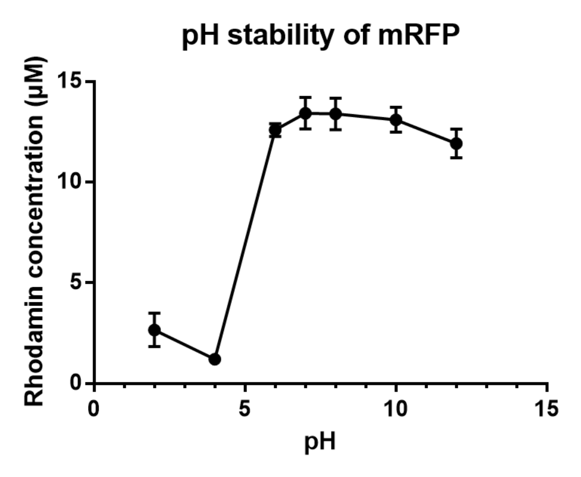
Summary: We measured the fluorescent signal of mRFP in buffers with different pH.
Documentation:
Charaterization of mRFP pH stabillity:

We grew C41 bacteria with parts BBa_J61002 in 2XYT for 24 hours. After purifying the mRFP by Ion Exchange Chromatography and Hydrophobic Interaction Chromatography, we measured the fluoresece (ex ,em ) of purified mRFP, which is diluted to 10µg/100µl (total 200µl) in triplicates, into different buffers (ranges from pH2 to pH12; Volume of mRFP:buffer = 1:1.8). To facilitate reproducibility of the experiment, we correlated the relative fluorescent intensity to an absolute fluorophore concentration by referring it to a standard curve of the fluorophores(Rhodamine) using the interlab study protocol. The result shows that the stability drops dramatically in pH condition below 6 and relatively stable in pH 6-10.

Fig.1 Vary pH attributed to different fluorescent intensity of RFP.

Table 1 Plate reader setting of fluorescent measurement

Measurement Type Fluorescence
Microplate name</td> COSTAR 96
Scan mode orbital averaging
Scan diameter [nm] 3
Excitation 550-20
Emission 605-40
Dichronic filter auto 572.5
Gain 500
Focal height [nm] 9

|};


Parts table

Protein data table for BioBrick BBa_E1010 automatically created by the BioBrick-AutoAnnotator version 1.0
Nucleotide sequence in RFC 10: (underlined part encodes the protein)
 ATGGCTTCC ... ACCGGTGCTTAATAACGCTGATAGTGCTAGTGTAGATCGC
 ORF from nucleotide position 1 to 675 (excluding stop-codon)
Amino acid sequence: (RFC 25 scars in shown in bold, other sequence features underlined; both given below)

101 
201 
MASSEDVIKEFMRFKVRMEGSVNGHEFEIEGEGEGRPYEGTQTAKLKVTKGGPLPFAWDILSPQFQYGSKAYVKHPADIPDYLKLSFPEGFKWERVMNFE
DGGVVTVTQDSSLQDGEFIYKVKLRGTNFPSDGPVMQKKTMGWEASTERMYPEDGALKGEIKMRLKLKDGGHYDAEVKTTYMAKKPVQLPGAYKTDIKLD
ITSHNEDYTIVEQYERAEGRHSTGA*
Sequence features: (with their position in the amino acid sequence, see the list of supported features)
None of the supported features appeared in the sequence
Amino acid composition:
Ala (A)12 (5.3%)
Arg (R)9 (4.0%)
Asn (N)4 (1.8%)
Asp (D)14 (6.2%)
Cys (C)0 (0.0%)
Gln (Q)8 (3.6%)
Glu (E)22 (9.8%)
Gly (G)23 (10.2%)
His (H)5 (2.2%)
Ile (I)9 (4.0%)
Leu (L)12 (5.3%)
Lys (K)22 (9.8%)
Met (M)9 (4.0%)
Phe (F)10 (4.4%)
Pro (P)12 (5.3%)
Ser (S)12 (5.3%)
Thr (T)14 (6.2%)
Trp (W)3 (1.3%)
Tyr (Y)11 (4.9%)
Val (V)14 (6.2%)
Amino acid counting
Total number:225
Positively charged (Arg+Lys):31 (13.8%)
Negatively charged (Asp+Glu):36 (16.0%)
Aromatic (Phe+His+Try+Tyr):29 (12.9%)
Biochemical parameters
Atomic composition:C1135H1749N299O347S9
Molecular mass [Da]:25423.7
Theoretical pI:5.65
Extinction coefficient at 280 nm [M-1 cm-1]:32890 / 32890 (all Cys red/ox)
Plot for hydrophobicity, charge, predicted secondary structure, solvent accessability, transmembrane helices and disulfid bridges 
Codon usage
Organism:E. coliB. subtilisS. cerevisiaeA. thalianaP. patensMammals
Codon quality (CAI):excellent (0.84)good (0.72)good (0.68)good (0.74)good (0.78)good (0.71)
Alignments (obtained from PredictProtein.org)
SwissProt:Q9U6Y8 (86% identity on 221 AAs), P83690 (63% identity on 215 AAs)
TrEML:Q5S3G8 (95% identity on 225 AAs), D0VWW2 (94% identity on 220 AAs)
PDB:2h5q (94% identity on 216 AAs), 2qlg (94% identity on 215 AAs)
Predictions (obtained from PredictProtein.org)
Subcellular Localization (reliability in brackets)
Archaea:secreted (100%)
Bacteria:cytosol (52%)
Eukarya:cytosol (20%)
Gene Ontology (reliability in brackets)
Molecular Function Ontology: -
Biological Process Ontology:GO:0018298 (40%), GO:0008218 (27%)
 
Predicted features:
Disulfid bridges: -
Transmembrane helices: -
The BioBrick-AutoAnnotator was created by TU-Munich 2013 iGEM team. For more information please see the documentation.
If you have any questions, comments or suggestions, please leave us a comment.


[edit]
Categories
//chassis/prokaryote/bsubtilis
//chassis/prokaryote/ecoli
//function/reporter
//function/reporter/fluorescence
//test
Parameters
abs
biology
colorRed
directionForward
emission607
emit607
excitation584
excite584
kegg
lum
proteinmRFP1
swisspro
tagNone