Difference between revisions of "Part:BBa J61101"
m |
|||
| Line 77: | Line 77: | ||
<h4>Evaluation of RBS strength in <i>Pseudomonas fluorescens</i></h4> | <h4>Evaluation of RBS strength in <i>Pseudomonas fluorescens</i></h4> | ||
<p>The RBS BBa_J61101 was tested in <i>P. fluorescens</i> and it's strength compared to two further ribosomal binding sites: <a href="https://parts.igem.org/Part:BBa_J61100">BBa_J61100</a> and <a href="https://parts.igem.org/Part:BBa_K4757003">BBa_K4757003</a></p> | <p>The RBS BBa_J61101 was tested in <i>P. fluorescens</i> and it's strength compared to two further ribosomal binding sites: <a href="https://parts.igem.org/Part:BBa_J61100">BBa_J61100</a> and <a href="https://parts.igem.org/Part:BBa_K4757003">BBa_K4757003</a></p> | ||
| − | <p>The parts strength was calculated by measuring the expression strength of the constitutively expressed fluorescence gene mKate2(<a href="https://parts.igem.org/Part:BBa_K4757000">BBa_K4757000</a> and calculating the fold change compared to the auto fluroescence of <i>P.fluorescens</i>.</p> | + | <p>The parts strength was calculated by measuring the expression strength of the constitutively expressed fluorescence gene mKate2(<a href="https://parts.igem.org/Part:BBa_K4757000">BBa_K4757000</a> and calculating the fold change compared to the auto fluroescence of <i>P. fluorescens</i>.</p> |
<img src="https://static.igem.wiki/teams/4757/wiki/fold-change-fluorescence-intensity-with-testsed-rbs.svg" alt"RBS_comp" width="300"> | <img src="https://static.igem.wiki/teams/4757/wiki/fold-change-fluorescence-intensity-with-testsed-rbs.svg" alt"RBS_comp" width="300"> | ||
<p display="table-caption" style="padding:10px" style="justif"><strong>Figure: Calculated fold change of fluorescence intensity of mKate2 with different ribosomal binding sites </strong><br>(RBS 1: BBa_J61100, RBS 2: BBa_J61101, RBS 3: synthetic de-novo RBS) compared to wild-type P. fluorescens. Measurements were taken in the early stationary phase. Values are presented as mean +/-SD. For statistical analysis groups were compared to wild-type P. fluorescens Reported significances were determined with ordinary One-way ANOVA with Dunn's method for multiple comparisons. *p < 0.05; **p < 0.01; ***p < 0.001; ****p < 0.0001. All differences in fold changes were significant (p < 0.001 for RBS 1 and 2. To reduce complexity only edge cases are shown.</p> | <p display="table-caption" style="padding:10px" style="justif"><strong>Figure: Calculated fold change of fluorescence intensity of mKate2 with different ribosomal binding sites </strong><br>(RBS 1: BBa_J61100, RBS 2: BBa_J61101, RBS 3: synthetic de-novo RBS) compared to wild-type P. fluorescens. Measurements were taken in the early stationary phase. Values are presented as mean +/-SD. For statistical analysis groups were compared to wild-type P. fluorescens Reported significances were determined with ordinary One-way ANOVA with Dunn's method for multiple comparisons. *p < 0.05; **p < 0.01; ***p < 0.001; ****p < 0.0001. All differences in fold changes were significant (p < 0.001 for RBS 1 and 2. To reduce complexity only edge cases are shown.</p> | ||
Revision as of 09:01, 12 October 2023
Ribosome Binding Site Family Member Parts J61100-J61150 are a family of similar ribosome binding site basic parts identified from a saturation mutagenic library.
 Library    TCTAGAGAAAGANNNGANNNACTAGT
 J61100     tctagaGAAAGAGGGGACAAactagt
 J61101     tctagaGAAAGACAGGACCCactagt
 J61102     tctagaGAAAGATCCGATGTactagt
 J61103     tctagaGAAAGATTAGACAAactagt
 J61104     tctagaGAAAGAAGGGACAGactagt
 J61105     tctagaGAAAGACATGACGTactagt
 J61106     tctagaGAAAGATAGGAGACactagt
 J61107     tctagaGAAAGAAGAGACTCactagt
 J61108     tctagaGAAAGACGAGATATactagt
 J61109     tctagaGAAAGACTGGAGACactagt
 J61110     tctagaGAAAGAGGCGAATTactagt
 J61111     tctagaGAAAGAGGCGATACactagt
 J61112     tctagaGAAAGAGGTGACATactagt
 J61113     tctagaGAAAGAGTGGAAAAactagt
 J61114     tctagaGAAAGATGAGAAGAactagt
 J61115     tctagaGAAAGAAGGGATACactagt
 J61116     tctagaGAAAGACATGAGGCactagt
 J61117     tctagaGAAAGACATGAGTTactagt
 J61118     tctagaGAAAGAGACGAATCactagt
 J61119     tctagaGAAAGATTTGATATactagt
 J61120     tctagaGAAAGACGCGAGAAactagt
 J61121     tctagaGAAAGAGACGAGTCactagt
 J61122     tctagaGAAAGAGAGGAGCCactagt
 J61123     tctagaGAAAGAGATGACTAactagt
 J61124     tctagaGAAAGAGCCGACATactagt
 J61125     tctagaGAAAGAGCCGAGTTactagt
 J61126     tctagaGAAAGAGGTGACTCactagt
 J61127     tctagaGAAAGAGTGGAACTactagt
 J61128     tctagaGAAAGATAGGACTCactagt
 J61129     tctagaGAAAGATTGGACGTactagt
 J61130     tctagaGAAAGAAACGACATactagt
 J61131     tctagaGAAAGAACCGAATTactagt
 J61132     tctagaGAAAGACAGGATTAactagt
 J61133     tctagaGAAAGACCCGAGACactagt
 J61134     tctagaGAAAGACCGGAAATactagt
 J61135     tctagaGAAAGACCGGAGACactagt
 J61136     tctagaGAAAGAGCTGAGCAactagt
 J61137     tctagaGAAAGAGTAGATCAactagt
 J61138     tctagaGAAAGATATGAATAactagt
 J61139     tctagaGAAAGATTAGAGTCactagt
These parts are present in plasmid pSB1A2, but there is also a constitutive promoter (J23100-derived) inserted into the XbaI site. So, for example, the EcoRI/PstI region of part J61100 reads:
 Biobrick 5'    XbaI                    J23100              XbaI    RBS Part     Biobrick 3'
 gaattcgcggccgcttctagaGTTGACGGCTAGCTCAGTCCTAGGTACAGTGCTAGCTtctagaGAAAGAGGGGACAAactagtagcggccgctgcag
This feature in no way prevents the use of these parts in standard Biobrick assembly. Normal prefix insertion into EcoRI/XbaI will delete this promoter element. Suffix insertion into SpeI/PstI will retain this promoter, but it can of course be removed later by a prefix insertion.
Note also that the base 5' to the SpeI site is allowed to float in these parts and is therefore rarely "T". The "G" downstream of the XbaI site obeys the standard. Because the database does not permit variation at this position, the predicted sequences of composite parts derived from these parts will be incorrect at this position.
More on this family of parts and their quantitative behavior is described here.
Add more about the biology of this part here
>Internal Priming Screening Characterization of BBa_J61101: Has no possible internal priming sites between this BioBrick part and the VF2 or the VR primer.
The 2018 Hawaii iGEM team evaluated the 40 most frequently used BioBricks and ran them through an internal priming screening process that we developed using the BLAST program tool. Out of the 40 BioBricks we evaluated, 10 of them showed possible internal priming of either the VF2 or VR primers and sometime even both. The data set has a range of sequence lengths from as small as 12 bases to as large as 1,210 bases. We experienced the issue of possible internal priming during the sequence verification process of our own BBa_K2574001 BioBrick and in the cloning process to express the part as a fusion protein. BBa_K2574001 is a composite part containing a VLP forming Gag protein sequence attached to a frequently used RFP part (BBa_E1010). We conducted a PCR amplification of the Gag-RFP insert using the VF2 and VR primers on the ligation product (pSB1C3 ligated to the Gag + RFP). This amplicon would serve as template for another PCR where we would add the NcoI and BamHI restriction enzyme sites through new primers for ligation into pET14b and subsequent induced expression. Despite gel confirming a rather large, approximately 2.1 kb insert band, our sequencing results with the VR primer and BamHI RFP reverse primer gave mixed results. Both should have displayed the end of the RFP, but the VR primer revealed the end of the Gag. Analysis of the VR primer on the Gag-RFP sequence revealed several sites where the VR primer could have annealed with ~9 - 12 bp of complementarity. Internal priming of forward and reverse primers can be detrimental to an iGEM project because you can never be sure if the desired construct was correctly inserted into the BioBrick plasmid without a successful sequence verification.
Usage and Biology
Group: Valencia_UPV iGEM 2018
Author: Adrián Requena Gutiérrez, Carolina Ropero
Summary: We have adapted the part to be able to assemble transcriptional units with the Golden Gate method. It also includes the BioBrick equivalent scar in the 3' extreme, so the insertion of this supplementary bases ensure the correct spacing for the coding sequence expression when assembled in a composite part.
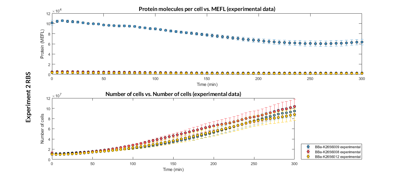
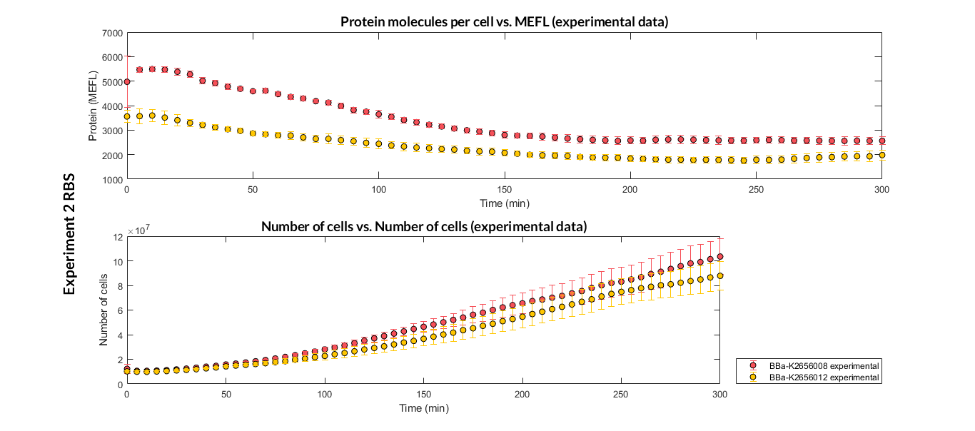
Characterization of the this part was performed with the transcriptional unit BBa_K2656104, which was used in a comparative RBS expression experiment with composite parts BBa_K265610 and BBa_K2656101. They all were assembled in a Golden Braid alpha1 plasmid using the same promoter, CDS and terminator.
By using this [http://2018.igem.org/Team:Valencia_UPV/Experiments#exp_protocol experimental protocol], we have obtained the parameters to valide our [http://2018.igem.org/Team:Valencia_UPV/Modeling#models constitutive model]and rationale choose its optimized values based on each RBS.
| Table 1. Optimized parameters for the BBa_K2656008 RBS. | |||
| Parameter | Value | ||
| Translation rate p | p = 0.02889 min-1 | ||
| Dilution rate μ | μ = 0.0118 min-1 | ||
We have also calculated the relative force between the different RBS, taking BBa_K2656009 strong RBS as a reference. It has been defined as the quotient between the values of the protein in equilibrium of the results of the simulation of one RBS and the reference RBS. Likewise, a ratio between p parameters of the different RBS parts and p parameter of the reference RBS has been calculated.
| Table 2. BBa_K2656008 relative strength and p ratio. | |||
| Parameter | Value | ||
| Relative strength | 0.032 | ||
| p parameter ratio (pRBS/pref) | 0.032 | ||
Sequence and Features
- 10COMPATIBLE WITH RFC[10]
- 12COMPATIBLE WITH RFC[12]
- 21COMPATIBLE WITH RFC[21]
- 23COMPATIBLE WITH RFC[23]
- 25COMPATIBLE WITH RFC[25]
- 1000COMPATIBLE WITH RFC[1000]
Team Warsaw 2010's measurement
RBS strength (relative to B0034): 22,70%
Team TU Delft 2010's measurements
RBS strength: 11.9%, standard deviation: 2.14%RBS strength is relative to B0034, obtained from an average of 12 measurements. Protein production rate is calculated using our production model
Measurements from 2023 team Heidelberg
Evaluation of RBS strength in Pseudomonas fluorescens
The RBS BBa_J61101 was tested in P. fluorescens and it's strength compared to two further ribosomal binding sites: BBa_J61100 and BBa_K4757003
The parts strength was calculated by measuring the expression strength of the constitutively expressed fluorescence gene mKate2(BBa_K4757000 and calculating the fold change compared to the auto fluroescence of P. fluorescens.

Figure: Calculated fold change of fluorescence intensity of mKate2 with different ribosomal binding sites
(RBS 1: BBa_J61100, RBS 2: BBa_J61101, RBS 3: synthetic de-novo RBS) compared to wild-type P. fluorescens. Measurements were taken in the early stationary phase. Values are presented as mean +/-SD. For statistical analysis groups were compared to wild-type P. fluorescens Reported significances were determined with ordinary One-way ANOVA with Dunn's method for multiple comparisons. *p < 0.05; **p < 0.01; ***p < 0.001; ****p < 0.0001. All differences in fold changes were significant (p < 0.001 for RBS 1 and 2. To reduce complexity only edge cases are shown.
Constructs with BBa_J61100 (RBS 1) showed minimal fold-change in fluorescence levels (0.73 +/- 0.316). The second RBS from the Anderson library (BBa_J61101, RBS 2) had a distinct increase in fold change compared to BBa_J61100 (18.15 +/- 3.21 compared to 0.73). The synthetic RBS (RBS 3) designed by the Salis-lab calculator (calculated for maximal expression strength for mKate2 mRNA) showed the strongest fluorescence (48.56 +/- 3.394).
Functional Parameters: Austin_UTexas
Burden Imposed by this Part:

Burden is the percent reduction in the growth rate of E. coli cells transformed with a plasmid containing this BioBrick (± values are 95% confidence limits). This BioBrick did not exhibit a burden that was significantly greater than zero (i.e., it appears to have little to no impact on growth). Therefore, users can depend on this part to remain stable for many bacterial cell divisions and in large culture volumes. Refer to any one of the BBa_K3174002 - BBa_K3174007 pages for more information on the methods, an explanation of the sources of burden, and other conclusions from a large-scale measurement project conducted by the 2019 Austin_UTexas team.
This functional parameter was added by the 2020 Austin_UTexas team.


