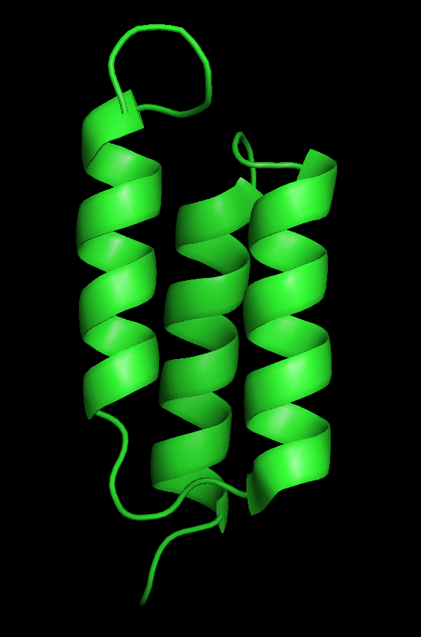
Part:BBa_K404302
Z-EGFR-1907
| [Z-EGFR-1907 | |
|---|---|
| BioBrick Nr. | BBa_K404302 |
| RFC standard | RFC 25 |
| Requirement | pSB1C3 |
| Source | |
| Submitted by | [http://2010.igem.org/Team:Freiburg_Bioware FreiGEM 2010] |
Usage in Biology
Affibodies are small (6 kDa), soluble high-affinity proteins. They are derived from the IgG-binding B domain of the Staphylococcal protein A, which was engineered to specifically bind to certain peptides or proteins. This so-called Z domain consists of an antiparallel three-helix bundle and is advantageous due to its proteolytic and thermodynamic stability, its good folding properties and the ease of production via recombinant bacteria (Nord et al. 1997). Affibodies can be used for example for tumor targeting (Wikman et al. 2004) and diagnostic imaging applications (Friedman et al. 2008)(Orlova et al. 2007). The ZEGFR:1907 Affibody was engineered to specifically bind the EGF receptor with an affinity of KD = 2.8 nM (Friedman et al. 2008).
The EGF receptor is overexpressed in certain types of tumors, e.g. in breast (Walker and Dearing 1999), lung (Hirsch et al. 2003) and bladder (Colquhoun and Mellon 2002) carcinomas, and is therefore a suitable target for cancer imaging or therapeutic applications. Because of their property to become internalized into the target cells with an efficiency of 19 – 24% within one hour – compared to 45% of the natural ligand EGF - the ZEGFR:1907 Affibody was chosen for therapeutic applications by the Freiburg iGEM Team 2010 (Göstring et al. 2010; Friedman et al. 2008).

Figure 1: Nucelotide sequence of designed ZEGFR:1907
Characterization
Specific targeting of tumor cells was, besides producing recombinant virus particles for therapeutical applications, one intention of the Virus Construction Kit provided by the iGEM team Freiburg_Bioware 2010.
For development of targeting strategies against EGF receptor (EGFR) over-expressing cancer cells, exhaustive literature search for engineering the surface of the Adeno-associated virus 2 (AAV2) was performed.
Besides insertion of targeting motifs into the viral protein 1 (VP1) open reading frame (ORF), we designed a method for fusing larger motifs to the N-terminus of VP2. It is expected that these peptides become located on the virus surface either by transit through the pores or by exposure during capsid assembly.
Additionally it is required to knock down the natural tropism of the virus towards its primary receptor heparan sulfate proteoglycan (HSPG) in order to prevent infection of healthy cells (Perabo et al. 2006). The binding motif consists of five amino-acids located on the capsid surface: R484/R487, K532, R585/587. (Trepel et al. 2009). The positively charged arginine residues interact with the HSPGs' negatively charged acid residues. Two point mutations (R585A and R588A) are sufficient to eliminate the heparin binding affinity of AAV2 (Opie et al. 2003). This function is also provided as Viral Brick by the Freiburg Virus Construction Kit (BBa_K404210, ViralBrick-587KO-Empty).
Transduction Efficacy by Flow Cytometry
For determination of transduction efficacy flow cytometry analysis was conducted. 250.000 AAV-293 cells were transfected with 1 µg total DNA. Different ratios of modified VP2/3 plasmids in respect to the Rep-VP13(587KO)_p5-TATAless plasmid (10:90, 25:75, 50:50) were co-transfected. 72 hours post transfection viruses were harvested and two different cell lines, HT1080 and A431, were transduced. By encapsulating mVenus coding sequence as gene of interest, the amount of transduced cells could be determined via flow cytometry.
|
HT1080 cells
A431 cells Figure2: Flow cytometry. Test of transduction efficiency with HT1080 and A431 cells by detecting mVenus expression from ZEGFR:1907_Middlelinker_VP2/3 virus particles (Transfection ratio: 50:50 in respect to Rep/Cap plasmid). A) Gating non transduced cells (control); subcellular debris and cellular aggregates can be distinguished from single cells by size, estimated via forward scatter (FS Lin) and granularity, estimated via side scatter (SS Lin). B) : Non transduced cells plotted against mVenus fluorescence (Analytical gate was set such that 1% or fewer of negative control cells fell within the positive region (R6). C) Gating transduced cells. D) Transduced cells plotted against mVenus fluorescence, R10 comprised transduced, mVenus expressing cells. E) Overlay of non-transduced (red) and transduced (green) cells plotted against mVenus fluorescence. |
Figure 3 overviews transduction efficacy of all ratio compositions.
|
Figure 3: Flow cytometry analysis. Transduced and therefore mVenus positive HT1080 and A431 cells, infected with virus particles consisting of different ratios of modified VP2/3 plasmids in respect to theRep-VP13(587KO)_p5-TATAless plasmid (10:90, 25:75, 50:50). |
Transduction of HT1080 cells revealed that all viral particles
remained infectious with efficacies up to 69 %, regardless of
which ratio
of pCMV_ZEGFR:1907_MiddleLinker_[AAV2]-VP23(ViralBrick-587KO-His-Tag)
plasmids
in respect to the Rep-VP13(587KO)_p5-TATAless plasmid was transfected.
In
general the amount of mVenus positive HT1080 cells decreased only
slightly when
harboring more modified VP2 subunits. A431 cells, which over express
EGF
receptor, were generally transduced with reduced efficacy. In
comparison to
HT1080 cells infection efficacy increased when inserting 50 %
modified VP2
plasmids. This indicates that the Affibody ZEGFR:1907 could
be
inserted into the AAV2 capsids without affecting virus assembly and
packaging.
Further on insertion of the Affibody into the capsid improved
infectivity,
clearly indicating that EGFR over expressing cells were successfully
targeted.
Infectious Titer by qPCR
We transfected 250.000 AAV-293 cells with 1 µg total DNA amount. Modified VP2/3 fusion plasmids were co-transfected to the Rep/Cap(VP2KO) plasmid in a 50:50 ratio. Viruses were harvested three days post transfection. The genomic titer was determined via qPCR by amplification of a specific sequence located in the CMV promoter of the vector plasmid (Table 1).
Table 1: Quantitative Real-Time PCR. Determination of the genomic titer. Data were corrected for negative control value.
|
Co-transfected Construct |
Ratio |
Genomic Titer /1ml Corrected For Negative Control |
|
Modified VP2/3 plasmid |
50:50 |
5,44E+08 |
|
Control: Rep/Cap |
100% |
1,55E+08 |
|
Control: Rep/Cap(587KO) |
100% |
5,39E+08 |
We investigated transduction of different cell lines. For this purpose 100.000 HT1080, HeLa or A431 cells were seeded and transduced with 50 µL virus stock and harvested 24 hours later. Infectious titers were determined via qPCR and normalized to the genomic titers (Fig. 4).

Figure 4: Infectious titers were determined for HT1080, HeLa and A431 cells. Control:Rep/Cap plasmid with and without HSPG knock down.
Figure 4 clearly demonstrates that HSPG affinity knocked
down
viruses have, in comparison to unmodified viruses, only slightly
infectious
properties towards HT1080 and HeLa cells. Both controls were nearly
were not
able to transduce A431 cells. In contrast infectivity was rescued by
integrating the Affibody ZEGFR:1907 into the virus capsids.
These
data emphasizes the functionality of the VP2 fusion construct for
specifically
targeting tumor cells for therapeutic or imaging applications.
Killing Tumor Cells: Time-Lapse
HT1080 and A431 cells were transduced with ZEGFR:1907_MiddleLinker_[AAV2]-VP23(ViralBrick-587KO-His-Tag) viruses packaged with the guanylate kinase fused to the thymidine kinase coding sequence (GMK-TK). A time series of pictures was started directly after adding 20 µM ganciclovir (Fig. 4).
|
Figure 4 a: Time-lapse. HT1080 (control) cells transduced with GMK-TK packaged viruses and treated with 20 µM ganciclovir A) 0 hours, B) 7 hours, C) 15 hours and D) 23 hours post transduction. |
|
Figure 4 b: Time-lapse. A431 cells transduced with GMK-TK packaged viruses and treated with 20 µM ganciclovir A) 0 hours, B) 7 hours, C) 15 hours and D) 23 hours post transduction. |
Virus and ganciclovir treatment only slightly affects the morphology of HT1080 cells. After 23 hours of incubation in 20 µM ganciclovir the cells had almost the same appearance as at time point zero (Fig. 4 a). In comparison to that A431 epidermoid carcinoma cells were efficiently killed after transduction and ganciclovir add-on: After 23 hours nearly all cells were lysed (Fig. 4 b).
These results clearly demonstrate that we were able to specifically target EGFR over-expressing tumor cells via capsid integrated Affibody and that these transduced cells were efficiently killed by expressing the GMK-TK which converted prodrug ganciclovir into its cell-toxic monophosphate.
References
Colquhoun, a J, and J K Mellon. 2002. Epidermal growth factor receptor and bladder cancer. Postgraduate medical journal 78, no. 924 (October): 584-9. doi:10.1136/pmj.78.924.584. http://www.pubmedcentral.nih.gov/articlerender.fcgi?artid=1742539&tool=pmcentrez&rendertype=abstract.
Friedman, Mikaela, Anna Orlova, Eva Johansson, Tove L J Eriksson, Ingmarie Höidén-Guthenberg, Vladimir Tolmachev, Fredrik Y Nilsson, and Stefan Ståhl. 2008. Directed evolution to low nanomolar affinity of a tumor-targeting epidermal growth factor receptor-binding affibody molecule. Journal of molecular biology 376, no. 5: 1388-402. doi:10.1016/j.jmb.2007.12.060. http://www.ncbi.nlm.nih.gov/pubmed/18207161.
Göstring, Lovisa, Ming Tsuey Chew, Anna Orlova, Ingmarie Höidén-guthenberg, Anders Wennborg, Jörgen Carlsson, and Fredrik Y Frejd. 2010. Quantification of internalization of EGFR-binding Affibody molecules: Methodological aspects. International Journal of Oncology 36, no. 4 (March): 757-763. doi:10.3892/ijo_00000551. http://www.spandidos-publications.com/ijo/36/4/757.
Hirsch, Fred R, Marileila Varella-Garcia, Paul a Bunn, Michael V Di Maria, Robert Veve, Roy M Bremmes, Anna E Barón, Chan Zeng, and Wilbur a Franklin. 2003. Epidermal growth factor receptor in non-small-cell lung carcinomas: correlation between gene copy number and protein expression and impact on prognosis. Journal of clinical oncology : official journal of the American Society of Clinical Oncology 21, no. 20 (October): 3798-807. doi:10.1200/JCO.2003.11.069. http://www.ncbi.nlm.nih.gov/pubmed/12953099.
Nord, K, E Gunneriusson, J Ringdahl, S Ståhl, M Uhlén, and P A Nygren. 1997. Binding proteins selected from combinatorial libraries of an alpha-helical bacterial receptor domain. Nature biotechnology 15, no. 8 (August): 772-7. doi:10.1038/nbt0897-772. http://www.ncbi.nlm.nih.gov/pubmed/9255793.
Orlova, Anna, Vladimir Tolmachev, Rikard Pehrson, Malin Lindborg, Thuy Tran, Mattias Sandström, Fredrik Y Nilsson, Anders Wennborg, Lars Abrahmsén, and Joachim Feldwisch. 2007. Synthetic affibody molecules: a novel class of affinity ligands for molecular imaging of HER2-expressing malignant tumors. Cancer research 67, no. 5 (March): 2178-86. doi:10.1158/0008-5472.CAN-06-2887. http://www.ncbi.nlm.nih.gov/pubmed/17332348.
Walker, R a, and S J Dearing. 1999. Expression of epidermal growth factor receptor mRNA and protein in primary breast carcinomas. Breast cancer research and treatment 53, no. 2 (January): 167-76. http://www.ncbi.nlm.nih.gov/pubmed/10326794.
Wikman, M, a-C Steffen, E Gunneriusson, V Tolmachev, G P Adams, J Carlsson, and S Ståhl. 2004. Selection and characterization of HER2/neu-binding affibody ligands. Protein engineering, design & selection : PEDS 17, no. 5 (May): 455-62. doi:10.1093/protein/gzh053. http://www.ncbi.nlm.nih.gov/pubmed/15208403.
Sequence and Features
- 10COMPATIBLE WITH RFC[10]
- 12COMPATIBLE WITH RFC[12]
- 21COMPATIBLE WITH RFC[21]
- 23COMPATIBLE WITH RFC[23]
- 25COMPATIBLE WITH RFC[25]
- 1000COMPATIBLE WITH RFC[1000]
//viral_vectors/aav/miscellaneous
| None |


