Designed by: Vanessa Kr�mer, Matthias Otto Group: iGEM18_Bielefeld-CeBiTec (2018-08-31)
Mutated Human Ferritin Heavy Chain
Short summary
The Human Ferritin Heavy Chain (HUHF) BBa_K2638999 was successfully cloned and expressed in Escherichia coli DH5 alpha. After protein purification HUHF was used to produce gold and silver nanoparticles which was ensured by examinations with the Transmission Electron Microscope and Energy-dispersive X-ray spectroscopy (EDX). Thus, we improved BBa_K1189019 which is not able to form gold and silver nanoparticles.
The Calgary 2013 iGEM team used the human ferritin wildtype (BBa_K1189019) as reporter protein for a test strip. They expressed the human ferritin heavy and light chain heterologous using Escherichia coli. In the cells, the ferritin produced its characteristic iron core, which was colored with the help of fenton chemistry to produce the prussian blue iron complex. Beside the function as reporter, the team mentioned the capability of ferritin to produce nanoparticles from other metal ions.
Figure 1: Ferritin is suitable for metal recycling, since it can form e.g. iron, silver and gold nanoparticles in its cavity.
The capability of human ferritin to bind different metal ions and form nanoparticles makes it suitable for the recycling of different valuable metal ions (Ensign et al., 2004; Domínguez-Vera et al., 2007). In addition, nanoparticles formed inside/by ferritin have advantages over nanoparticles produced by conventional methods. On the one hand the ferritin encapsulated nanoparticles are water soluble due to the protein shell. On the other hand the maximal inner diameter of ferritin of 8 nm causes a upper size restriction of the nanoparticles inside the ferritin (Butts et al., 2008). This restriction is desirable for various applications (Castro et al., 2014).
The wildtype ferritin has reactive amino acids on the outside and inside of the protein shell, causing nanoparticle synthesis at both surfaces. An optimization of the wildtype human ferritin towards a nanoparticle syntheses mainly in the interior can therefore favor a unified production of different nanoparticles (Butts et al., 2008).
Improved Human Ferritin: BBa_K2638999
To improve the ferritins capability to direct metal ions to its inside and to increase its ability to form gold and silver nanoparticles, we constructed a mutated version of the human ferritin heavy chain (HUHF) (BBa_K2638999). Following Christopher A. Butts et al. (2008) we removed reactive cysteine and histidine residues from exterior of the HUHF and added additional cysteine residues at the interior. This way the production of nanoparticles at the exterior surface is prevented or at least decreased.
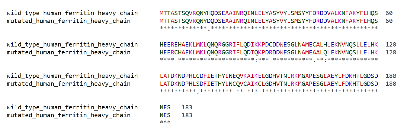
Figure 2: Alignment of the protein sequences of the wildtype and the mutated human ferritin heavy chain. The Alignment was produced with Clustal Omega (Goujon et al., 2010, Sievers et al., 2011).
In Figure 2 the amino acid sequence alignment of the wildtype human ferritin and the mutated human ferritin is shown. The exterior residues C91R, C103A, C131S, H14D and H106Q, and the interior residues E65C, E141C, E148C, K87Q and K144C were mutated. The mutations have no influence on the structure of the ferritin as shown in Figure 3.
Figure 3: Protein structures of the wildtype human ferritin (A, RCSB ID 4oYN) and the mutated human ferritin (B, RCSB ID 3ES3). Despite the mutations of ten amino acids the ferritin retains its shape. The protein structeres were generated with Chimera (Pettersen et al., 2004).
We ordered the CDS for the Human Ferritin Heavy Chain (HUHF) as gBlock from IDT. Due to the complications of the synthesis of HUHF l, we had to split the protein into three parts with 50 basepairs overlapping sequences to each other and to the backbone pSB1C3. This enables combined cloning of all three HUHF-gBlocks by Gibson Assembly into the plasmid backbone of pSB1C3, thus creating the basic part BBa_K2638999 .
For expression of HUHF we cloned an araBAD promoter (BBa_I0500) and a ribosome-binding site (RBS) (BBa_R0030) upstream of the CDS via BioBrick assembly.
HUHF was expressed in E. coli DH5α. Therefore, 50 mL LB cultures in shaking flasks were inoculated from an overnight culture to achieve an OD600 of 0.1. The cultures were cultivated at 37 °C and 140 rounds per minute (rpm). After growing to an OD600 of 0.6-0.8, HUHF expression was induced with 1 % L-arabinose. After induction the flasks were incubated at 28 °C and 140 rpm for six hours. Samples were taken hourly for sodium dodecyl sulfate polyacrylamide gel electrophoresis (SDS-PAGE).
Moreover, the samples were further purified. Therefore, the protocol of Dr. Jon Marles Wright has been applied. The samples were pelleted (13000g, 1 min), the media was discarded and cell pellets were resuspended in buffer A (50 mM tris pH 8, 1 mM DTT, 0.1 mM ethylenediaminetetraacetic acid, 20 mM mannitol). Afterwards the cells were lysed by sonication, centrifuged (10000 g, 20min), heated to 80 °C for 10 minutes, put on ice for 10 minutes and centrifuged (10000 g, 10 min) again. Purified HUHF was located in the supernatant.
HUHF purified by this workflow was analyzed with a SDS-PAGE (Fig. 1). Clearly visible bands have been recognizable at about 23 kDa, the expected hight for HUF.
Figure 4: SDS-PAGE of purified Human Ferritin Heavy Chain (BBa_K2683999). HUHF under control of the araBAD promoter (BBa_I0500) was expressed in E. coli DH5 alpha and afterwards purified. Shown are samples were expression was induced with 1 % arabinose (third, sixth ninth and twelfth lane) and controls without induction (second, fifth, eighth and eleventh lane) as well as empty-vector controls (pSB1C3 without insert, first, fourth, seventh and tenth lane).
The SDS-PAGE of the purified HUHF showed intense bands at about 25 kDa, fitting to the theoretical molecular weight of HUHF of 24.087 kDa. To ensure the identity of the HUHF the band was cut out of the SDS-PAGE and prepared for matrix assisted laser desorption ionisation with time of flight analysis (MALDI-TOF) measurements. These show distinctive peaks for the HUHF. In addition, tandem mass spectrometry measurements show peptides specific for HUHF, thereby confirming HUHF expression as well as a successful purification workflow. Besides, measurements reveal that the HUHF has been expressed.
Figure 5: Results of MALDI-TOF measurements. The peptide fragments show a recognizable pattern for the HUHF.
In Figure 5 the specific peptide fragments pattern of the HUHF can be seen. The samples were digested with trypsin prior MALDI-TOF. Per tandem mass spectrometry the peak with the m/z = 1345.649 was further examined (Figure 6).Thus, all results confirm that the measured sample contains HUHF.
Figure 6: Results of the tandem mass spectrometry of the peak 1345 of HUHF.
The purified HUHF was used to produce gold and silver nanoparticles. Therefore, HUHF samples were prepared by removing the ferritin bound Fe3+ ions. By applying the (name of the protocol) protocol, the Fe3+ ions have been reduced to Fe2+ ions. After the iron ions were removed, AuHCl4 and AgNO3 solutions were added as stated in the (name of the protocol) protocols. The HUHF with the AuHCl4 solution was incubated for 1.5 hours whereas the AgNO3 solution was incubated for 18 hours while being illuminated by a 60 watts lamp. Afterwards the samples were centrifuged (10.000 g, 10 min) and further purified with a 100 kDa protein columns to remove denaturated HUHF.
For demonstration of nanoparticle formation and determination of nanoparticle composition the samples were examined using a transmission electron microscope (TEM).
Analysis workflow: The TEM images of the nanoparticles were automatically analyzed in ImageJ. The particles were separated from the background by k-means clustering. Afterwards touching particles were segmented by watershed segmentation. The processed image was binarized and a particle analysis was performed.
Figure 7: Automatic identification of 147 silver nanoparticles in the wildtype human ferritin sample (BBa_K1189019).
Figure 7 shows a TEM image with 147 identified silver nanoparticles produced by the wild type human ferritin (BBa_K1189019). The particles are between 24.5 and 1597.8 nm in size with one very big particle with a size of 7272.3 nm, which seems to consist in many agglutinated silver nanoparticles. No particle was found in the expected size of about 8 nm.
Figure 8: Automatic identification of 708 silver nanoparticles in the gold silver mutant ferritin sample (BBa_K2638999). 431 (60.8%) of the nanoparticles had a mean diameter of 8 nm or less.
Figure 8 shows a TEM image with 708 identified silver nanoparticles produced by the gold silver mutant ferritin sample (BBa_K2638999). The particles have a size between 1.8 and 34.8 nm. 120 of the silver nanoparticles (16.9 %) are exactly in the expected size of 7 to 9 nm which indicates that at least all of these particles are produced by our improved ferritin (BBa_K2638999).
Figure 9: The silver nanoparticles in our gold silver mutant ferritin (BBa_K2638999) with a mean diameter of 8.2 nm were significant smaller than the nanoparticles of the wildtype human ferritin (BBa_K1189019) with a mean diameter of 531.8 nm.
The direct comparison of our new gold silver mutant ferritin (BBa_K2638999) and the old wild type human ferritin (BBa_K1189019) in figure 9 shows that our improved enzyme produces nearly five times more silver nanoparticles which are 98.5 % smaller than the silver nanoparticles produced by the wild type ferritin. This proves that the new ferritin enzyme is much more suitable for producing silver nanoparticles than the wild type version.
Figure 10: Automatic identification of 2 gold nanoparticles (13 and 10 nm) in the wildtype human ferritin sample (BBa_K1189019).
Figure 11: Automatic identification of 2 gold nanoparticles (7 and 9 nm) in the gold silver mutant ferritin sample (BBa_K2638999). These nanoparticles are approximately 30 % smaller than the nanoparticles produced by the wildtype ferritin.
Outlook
Nanoparticles produced with ferritin can be used in various apllications (Figure 12). As example, they can be directly used inside the ferritin cage for moleculer imaging (Wang et al., 2017b). When extracted, they can be used as antibacterial agent, in particular silver nanoparticles (Wang et al., 2017a), as biosensor (Castro et al., 2014) or they can be printed and melted to produce electronic circuits (Ummartyotin et al., 2012). In particular, we have dealt with the printing of electronics in our project.
Figure 12: Possible applications of nanoparticles produced with ferritin.
Molecular graphics and analyses performed with UCSF Chimera, developed by the Resource for Biocomputing, Visualization, and Informatics at the University of California, San Francisco, with support from NIH P41-GM103311. Butts, C.A., Swift, J., Kang, S., Di Costanzo, L., Christianson, D.W., Saven, J.G., and Dmochowski, I.J. (2008).. Directing Noble Metal Ion Chemistry within a Designed Ferritin Protein † , ‡. Biochemistry 47: 12729–12739.
Castro, L., Blázquez, M.L., Muñoz, J., González, F., and Ballester, A. (2014).. Mechanism and Applications of Metal Nanoparticles Prepared by Bio-Mediated Process. Rev. Adv. Sci. Eng. 3.
Ensign, D., Young, M., and Douglas, T. (2004).. Photocatalytic synthesis of copper colloids from CuII by the ferrihydrite core of ferritin. Inorg. Chem. 43: 3441–3446.
Goujon, M., McWilliam, H., Li, W., Valentin, F., Squizzato, S., Paern, J., and Lopez, R. (2010).. A new bioinformatics analysis tools framework at EMBL-EBI. Nucleic Acids Res. 38: W695-699.
Pettersen, E.F., Goddard, T.D., Huang, C.C., Couch, G.S., Greenblatt, D.M., Meng, E.C., and Ferrin, T.E. (2004).UCSF Chimera--a visualization system for exploratory research and analysis. J Comput Chem 25: 1605–1612.
Sievers, F., Wilm, A., Dineen, D., Gibson, T.J., Karplus, K., Li, W., Lopez, R., McWilliam, H., Remmert, M., Söding, J., Thompson, J.D., and Higgins, D.G. (2011). Fast, scalable generation of high-quality protein multiple sequence alignments using Clustal Omega. Mol. Syst. Biol. 7: 539.
Ummartyotin, S., Bunnak, N., Juntaro, J., Sain, M., and Manuspiya, H. (2012). . DSynthesis of colloidal silver nanoparticles for printed electronics. /data/revues/16310748/v15i6/S1631074812000549/.
Wang, L., Hu, C., and Shao, L. (2017a).. The antimicrobial activity of nanoparticles: present situation and prospects for the future. Int. J. Nanomedicine 12: 1227–1249.
Wang, Z., Gao, H., Zhang, Y., Liu, G., Niu, G., and Chen, X. (2017b).. Functional ferritin nanoparticles for biomedical applications. Front. Chem. Sci. Eng. 11: 633–646.