Part:BBa_K2477013
Copper Biosensor
This part constitutively expresses the CueR repressor. This repressor then inhibits the expression of RFP from the downstream PcopA promoter. In the presence of copper, the repression of CueR is inhibited leading to greater RFP expression. Fluorescence of this construct has been shown to increase with increasing copper concentration in both a qualitative and quantitative manner.

Usage and Biology
This part utilizes machinery from the CueR copper regulon system of E. coli. This system has been extensively characterized and tested throughout the literature and across many iGEM teams. In its native setting, the CueR repressor acts to inhibit transcription of the copA and cueO genes which act as a copper efflux pump and copper oxidase respectively (1). In the presence of copper, the regulatory ability of CueR is inhibited. Hence, copA and cueO are able to expressed. The protein products of these genes act to decrease copper concentration within the cell. For this part, the CueR system was adapted for use within our biosensor part. Rather than expressing copA in the presence of copper, our system instead expresses RFP.
Characterization
For initial testing of this part, qualitative tests were conducted by inducing RFP expression using varying concentrations of copper in overnight cultures. Glycerol stocks of XL1-Blue cells transformed with the copper detection plasmid were used to inoculate 4 culture tubes containing 5 mL of LB broth. The appropriate antibiotic was added and then the cultures were induced to a final copper (II) sulfate concentration of 0 µM, 5 µM, 100 µM, or 500 µM. After incubation with shaking overnight, these cultures were spun down in a centrifuge and compared qualitatively. Clear differences in the color of the cell pellets could be seen between the 0 µM, 100 µM, and 500 µM groups. The 5 µM group appeared to be about the same coloration as the 0 µM group.

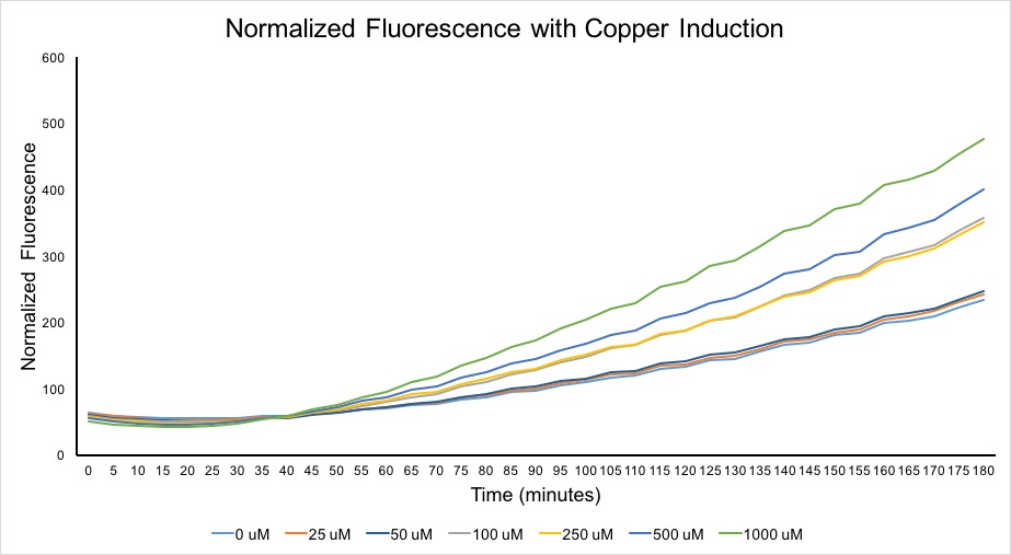
Following the initial test, a more quantitative experiment was performed. XL1-Blue cells containing the copper biosensor were grown overnight without induction to maximum culture density. Approximated 1 mL of this culture was then used to inoculate 20 mL of fresh LB in a 50 mL Falcon tube. This new culture was grown at 37 C with shaking until it reached an OD600 above .4. 1 mL samples of this culture were then removed and induced with copper (II) sulfate to final concentrations of 0 µM, 25 µM, 50 µM, 100, µM, 250 µM, 500 µM, and 1,000 µM. Aliquots of each induced sample were then loaded into 5 wells of a 96 well plate. This plate was placed into a shaking/incubating plate reader set at 37 C and measured for fluorescence (584 nm excitation 612 nm emission) every 5 minutes for 180 minutes in total. OD600 readings were taken at the beginning and end of this time course and a linear regression was used to approximate the OD600 for each time point. The OD600 data was used to normalize the fluorescence data in order to generate the graph below.
This graph shows clear differences in terms of the expression profiles of the different experimental groups. While the cells induced with 25 µM and 50 µM copper show little to no difference from the non-induced sample, the other induction groups show a clear upward progression in terms of fluorescence against increasing copper concentration. Overall, this figure as well as the qualitative testing show that our copper biosensor can reliably detect copper concentrations greater than or equal to 100 uM.
Potential Applications
From our research and after visiting the Washington Suburban Sanitation Commission Patuxent Water Treatment Plant, we found that normal drinking water typically contains around 0.01 mg/L copper. The EPA limit set on copper in drinking water is meanwhile 1.3 mg/L. When converted to µM, typical drinking water copper levels are approximately 0.2 µM whereas the EPA limit is set at approximately 20 µM. Since our biosensor sensitivity is limited to 100 µM, detection of copper levels above the EPA limit would require water samples to be concentrated prior to any form of induction. While this would not be the most convenient in terms of actually testing water for copper contamination, the functionality of this part has served our team well in terms of helping to educate high school students and the next generation of synthetic biologists. This part could potentially be used by future iGEM teams in order to facilitate discussion with the general public about the problem solving ability of synthetic biology.
Collaborative Efforts
This part was used as part of a collaboration with the William and Mary iGEM team. They added protein degradation tags onto the end of the RFP gene in order to incorporate it for use alongside their mf-Lon protease expression system so as to achieve control over gene expression speed. To read about these efforts, please reference our collaboration page or William and Mary's collaboration page (linked below). Alternatively, take a look at the parts they submitted that were derived from this part. The part numbers for the copper biosensor tagged with varying protein degradation tags are BBa_K2333437, BBa_K2333438, BBa_K2333439, BBa_K2333440, BBa_K2333441, and BBa_K2333442.
Link to UMaryland iGEM Collaboration Page: http://2017.igem.org/Team:UMaryland?target=collaborations
Link to William and Mary iGEM Collaboration Page: http://2017.igem.org/Team:William_and_Mary/Collaborations
References
1. Yamamoto K, Ishihama A. Transcriptional response of Escherichia coli to external copper. Molecular Microbiology. 2005;56(1):215-227. doi:10.1111/j.1365-2958.2005.04532.x.
Sequence and Features
- 10COMPATIBLE WITH RFC[10]
- 12INCOMPATIBLE WITH RFC[12]Illegal NheI site found at 7
Illegal NheI site found at 30 - 21COMPATIBLE WITH RFC[21]
- 23COMPATIBLE WITH RFC[23]
- 25INCOMPATIBLE WITH RFC[25]Illegal AgeI site found at 1237
Illegal AgeI site found at 1349 - 1000COMPATIBLE WITH RFC[1000]
| None |

