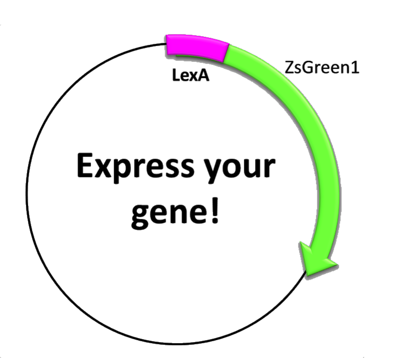
Part:BBa_K763004
pLexA + Gene encoding ZsGreen1
This construction consists of three elements:
- The transcription factor-binding site inside the promoter
- The repressor-binding site inside the promoter and
- the coding sequence, which contains a synthetic fluorescent (green) protein. The promoter was taken from the recA gene of E. coli, which participates in the orchestrated bacterial SOS-response [1] that involves more than forty independent SOS genes, most of which encode protein sengaged in protection, repairing and replication regulate our construction.
When is the protein synthesized? In order to obtain the green fluorescent protein one condition should be met. Since there is a repressor, lexA, that blocks any possible transcription, it is needed to UV-irradiate the cells to break down lexA and habilitate transcription.
The molecular mechanism underlying this phenomenon is as follows: when a bacterium undergoes severe damage it uses this repair system for maintaining a functional (double stranded) DNA able to duplicate. The damage can be so harmful that it can generate ruptures, dimers, bases modification, etc., therefore the bacteria releases RecA resulting in polymerized and filamentous structures which bind to and stabilize DNA [2], letting some error-prone DNA polymerases (translesion synthesis) to come and copy, randomly, the DNA. Solving the problem this way provokes several mutations in the bacteria's genome but it will stay alive because it maintains a functional DNA. Activation of the polymerases IV and V only occurs in advance stages of the response.The system comprises a negative regulator of the SOS response (LexA) and a positive regulator of the SOS response (RecA). The idea is that UV irradiation promotes LexA autocatalytic activity and RecA is activated. Then, LexA will not be able to bind to the promoters (SOS boxes or LexA boxes) and transcription will start (here is our construction with the protein we want). The process is relatively slow, reaching higher values of expression more than one hour later [3].
In our construction, we have picked out the LexA-binding site inside RecA promoter and then we have inserted a GFP sequence. Then, after irradiating we can see fluorescence… E. coli has accomplished our order!
How did we deal with this construction? We designed a UV-irradiation controlling device called Tordeitor 3000, so we could emit UV (254nm) without risk. After putting the plates, we irradiate different times in order to expose E. coli to different energies [4,5,6].
Characterization
In order to test our UV-induced construction, the E. coli strain carrying the ZsGreen1 gene under the control of the lexA promoter was grown on LBA (LB broth supplemented with ampicillin) until an OD value of 0.8. Then, cells were pelleted, resuspended in fresh medium, and 100 uL were spread in agar plates. Once the medium was completely absorbed by the agar (to avoid UV absorption by medium), cells were irradiated inside the Tordeitor (the device we specially designed for this experiment) with different amounts of UV light (lambda=254 nm). To fine tune UV dose, we used a shutter (similar to those used in cameras) able to produce UV pulses of less than 1 second of duration. After that, cells were harvested from the agar plate with SOC medium supplemented with ampicillin. Finally, OD and fluorescence intensity were measured at different times. An aliquot of the same culture that was not irradiated was used as control.
As we can see in the graph below, UV exposure does induce the expression of ZsGreen1. Furthermore, longer exposures result in higher levels of protein expression (higher fluorescence intensity was observed, as expected according to the molecular mechanism of lexA promoter), although lethal effects were observed when doses about 5 seconds were applied.
 |
 |
|
| Figure 9. Fluorescence intensity (FI) normalized by the optical density (OD) of cultures that were subjected to UV exposures of different duration in comparison to a control culture (not exposed). Measures were taken 70 min after the exposure. | Figure 10. Fluorescence intensity (FI) normalized by the optical density (OD) of cultures that were subjected to UV exposures of different duration in comparison to a control culture (not exposed). Measures were taken 90 min after the exposure. |
Sequence and Features
- 10COMPATIBLE WITH RFC[10]
- 12COMPATIBLE WITH RFC[12]
- 21COMPATIBLE WITH RFC[21]
- 23COMPATIBLE WITH RFC[23]
- 25COMPATIBLE WITH RFC[25]
- 1000COMPATIBLE WITH RFC[1000]
| None |

