Part:BBa_K4387994
Monovalent Anti-Tumour Necrosis Factor Nanobody (VHH#12B)
Single domain antibodies, also called VHH or Nanobody®, only consist of a single variable domain, which is able to bind to a specific antigen. The small size of nanobodies (15 - 20kDa) compared to antibodies, gives them special abilities, which are hard to reach with conventional antibodies. They can locally penetrate barriers (such as tissues) more easily and can withstand extreme environmental conditions, such as high temperatures and low pH. [1] They show high affinity and stability, and recombinant expression has revolutionized the biotechnology field. Nanobodies have already been discovered in camelid animals back in the 90's. Usage of these nanobodies in the clinic often requires an additional step called "humanization" in order to reduce unwanted immunological reactions upon administration. This step describes the exchange of one or a few specific amino acids that are recognized as "foreign" by the human immune system. [3] Still today, camelid animals are infected with the antigen of choice and effective nanobodies are obtained from their blood. However, new manufacturing technologies have been developed, allowing the screening of new candidates by using naive or synthetic libraries in combination with phage and ribosome display. The usage of synthetic libraries results in the generation of so called "sybodies". [2] Generating new nanobodies against a target is a time-consuming process with several selection steps. Therefore, we used the amino acid sequences for specific anti-tumour necrosis factor (TNFα) nanobodies from a patent and converted them to DNA sequences. [3] The patent contains a variety of TNFα-binding nanobodies. We selected 3 candidates (VHH#2B, VHH#3E and VHH#12B) based on their humanization characteristics. The patent described a complete humanization protocol for nanobody VHH#3E which we applied. The candidates VHH#2B and VHH#12B were described as already suitable for clinical applications due to their >90% amino acid sequence homology to human VH framework regions and are therefore possibly "safe" for direct administration to patients. The nanobody described below is the monovalent candidate VHH#12B.
Sequence and Features
- 10COMPATIBLE WITH RFC[10]
- 12COMPATIBLE WITH RFC[12]
- 21COMPATIBLE WITH RFC[21]
- 23COMPATIBLE WITH RFC[23]
- 25COMPATIBLE WITH RFC[25]
- 1000COMPATIBLE WITH RFC[1000]
Characterization
Purification of nanobodies
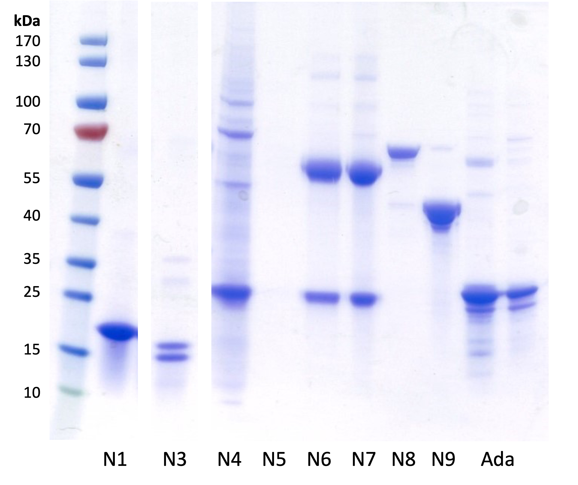
To show the TNFα-binding capacity of the nanobody and the inhibitory effect, we first inserted the nanobody sequence into the expression vector pSBinit by FX-cloning using the restriction enzyme SapI. The common lab strain E. coli MC1061 was transformed with the plasmid and nanobody expression was induced with arabinose. The nanobodies were extracted via periplasmic extraction (for monovalent constructs) or whole cell lysis (for bivalent constructs). Afterwards, they were purified via immobilized metal anion chromatography (IMAC). To insure, that the desired product was produced, we ran a native PAGE (Polyacrylamide Gel Electrophoresis) on the purified fractions (Figure 1). The sample labeled as N3 in figure 1 shows the monovalent nanobody VHH#12B. The obtained band shows the correct size of approximately 20 kDa.
ELISA
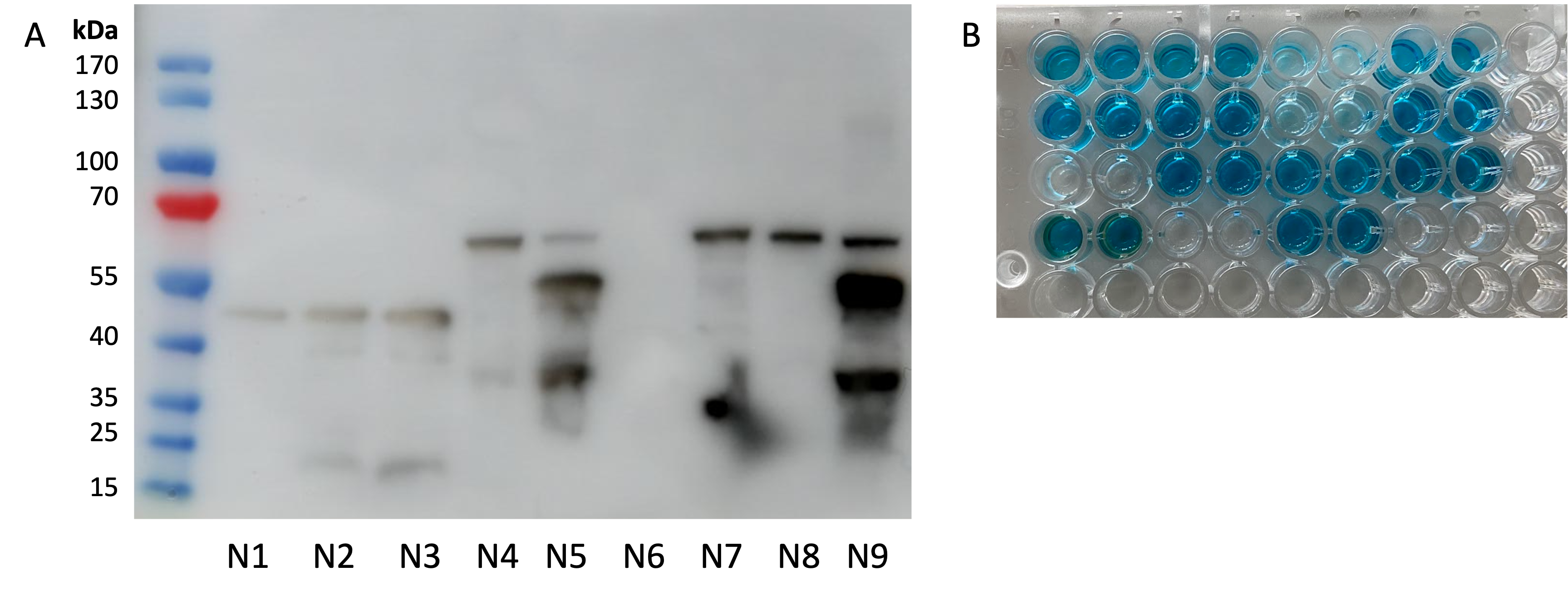
To proof the binding capacity of bacterially expressed anti-TNFα nanobodies, we conducted an ELISA with all purified monovalent and bivalent candidates (Figure 2). We could show that all candidates successfully bind TNFα. Adalimumab is a monoclonal antibody that is already used in the clinics for anti-TNFα therapies in IBD patients. It served as a positive control (wells D3-4). A random sybody against a membrane protein was used as a negative control (wells A1-2). Successful binding of nanobody VHH#12B is seen in wells B3-4.
Cell Assay
Lastly, we wanted to see if the nanobodies show an observable inhibitory effect on the immune response of human monocytes. We used the human monocytic cell line THP-1 and incubated the cells firstly with the nanobody candidates and Adalimumab. Afterwards, they were stimulated for 24 hours with different doses of TNFα, triggering their immune response. Successful inhibition of TNFα by the nanobody or Adalimumab is expected to reduce the inflammatory response of the monocytes. To quantify this, we isolated the cells’ RNA and performed a reverse transcription. We then compared the IL-1ß expression levels to the house-keeping gene GAPDH by quantitative real time PCR (qRT PCR). IL-1ß is a major inflammation mediator and a good marker for the intensity of the monocytic immune response that we induced with TNFα. Figure 3 shows the overall performance of the monovalent candidate VHH#12B. Adalimumab served as a positive control.
Figure 3 shows that TNFα alone induces a significant expression of IL-1β. In comparison, the tested monovalent nanobody could reduce the inflammatory response by up to 4-fold.
Secretion of the Nanobody with the Hemolysin A Secretion System
The hemolysin A secretion machinery is a one-step secretion system (T1SS), originally isolated from uropathogenic E. coli strains. [4] It comprises three main peptides, the inner membrane proteins HlyB and HlyD, and the outer membrane protein TolC. Together, these three proteins build a continuous channel through which originally the HlyA toxin is secreted in a one-step manner. Scientists have identified the secretion signal and were able to secrete various proteins of different sizes with this secretion machinery. [4]

The secretion of this monovalent nanobody with the HlyA secretion system (BBa_K4387987) is characterized in the respective composite part BBa_K4387984. We were able to show with a Western blot and ELISA that the common lab strain E. coli MC1061 (figure 4) and the probiotic strain E. coli Nissle 1917 are both able to secrete functional anti-TNFα nanobodies.
Additionally, we characterized the secretion of the monovalent nanobody VHH#2B with another inducible promoter, pNorVβ (BBa_K4387000). This alternative promoter senses nitric oxide (NO) and as a response to its binding, expresses the nanobody VHH#2B. This data is found in the respective composite part BBa_K4387978.
References
- [1] Harmsen, M.M., De Haard, H.J. Properties, production, and applications of camelid single-domain antibody fragments. Appl Microbiol Biotechnol 77, 13–22 (2007). https://doi.org/10.1007/s00253-007-1142-2
- [2] Zimmermann, I., Egloff, P., Hutter, C.A.J. et al. Generation of synthetic nanobodies against delicate proteins. Nat Protoc 15, 1707–1741 (2020). https://doi.org/10.1038/s41596-020-0304-x
- [3] Silence, Karen, Lauwereys, Marc, De Haard, Hans, et al. "Single domain antibodies directed against tumour necrosis factor-alpha and uses therefor", Int. Publication Number: WO 2004/041862 A2, 21 May 2004
- [4] Ruano-Gallego, D., Fraile, S., Gutierrez, C. et al. Screening and purification of nanobodies from E. coli culture supernatants using the hemolysin secretion system. Microb Cell Fact 18, 47 (2019). https://doi.org/10.1186/s12934-019-1094-0
| None |


