Part:BBa_K1739002
Sequence coding for Sup35 with a N-terminal CsgA signal sequence for amyloid export This part design includes the bipartite csgA signal sequence that targets the protein to the Sec-export pathway and subsequently to the Curli export pathway via interaction with csgG (Sivanathan and Hochschild, 2012; Sivanathan and Hochschild, 2013). Sup35NM is derived from the yeast prion protein Sup35p from S. cerevisiae (bakers yeast) and excludes the C-terminal domain with the N-terminal domain allowing self-assembly of functional amyloid (Frederick et al., 2014; Glover et al. 1997). This has previously been discussed by Tessier and Lindquist (2009) who show that two beta-sheets bond together in a self-complimenting ‘steric zipper’ that excludes water, leaving a highly stable parallel beta-sheet with one molecule every 4.7 Angstroms. The particular advantage of using Sup35NM is that in its native state Sup35p has two functional domains, the N and C terminal, separated by the highly charged M domain (Frederick et al., 2014; Glover et al. 1997; Wickner et al., 2007) allowing the fusion of a new functional domain. This part has been inserted into the pSB1C3 backbone and uses the promoter BBa_J23104.
Sequence and Features
- 10COMPATIBLE WITH RFC[10]
- 12INCOMPATIBLE WITH RFC[12]Illegal NheI site found at 7
Illegal NheI site found at 30 - 21COMPATIBLE WITH RFC[21]
- 23COMPATIBLE WITH RFC[23]
- 25COMPATIBLE WITH RFC[25]
- 1000INCOMPATIBLE WITH RFC[1000]Illegal SapI.rc site found at 718
Validation
Plasmid
To validate this part, it was first analyzed by a diagnostic restriction digest using EcoRI and PstI followed by agarose gel electrophoresis. We carried out a diagnostic restriction digest using ECORI and PSTI. These enzymes cleave pSBIC3 into a fragment of 2029bp, whereas our insert is 1020bp. We used the gel to check the sizes of our fragments against the Invitrogen 1kB plus DNA marker (see Fig 1.). We were therefore able to confirm that we had produced the correct part.
Congo Red Plate Assay
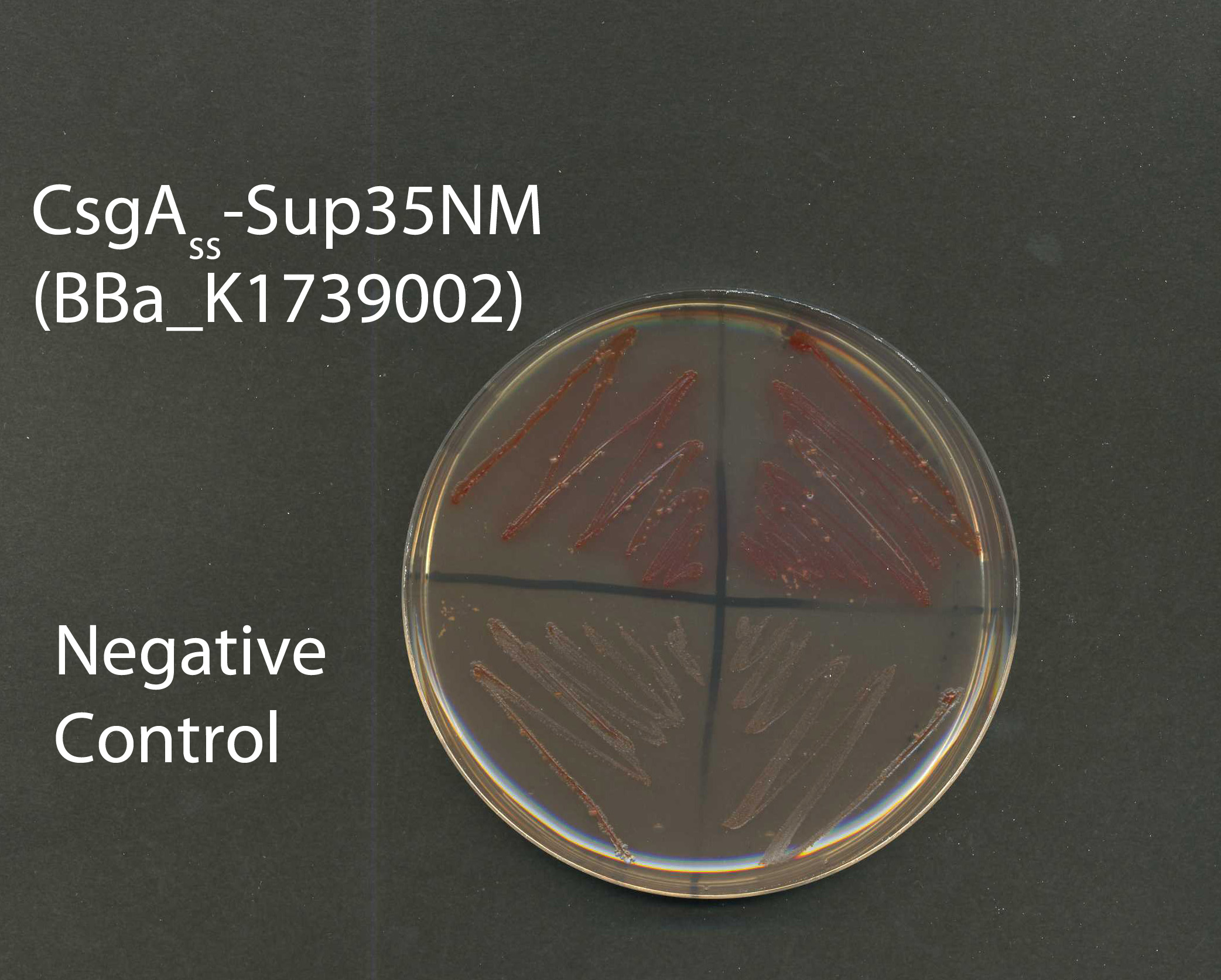
The protein this BioBrick is coding for has been validated using a Congo Red agar plate assay, which is an assay that confirms the presence of self-assembled amyloid. Our fusion protein was expressed and exported by VS45 E.coli cells. The Congo red plates were compared to a negative control strain, VS45 with pVS105. pVS105 contains CsgAss with Sup35M, this protein will not produce a protein product that self assembles into amyloid. The results (shown in Fig.2) confirmed that our protein self-assembled into amyloid nano-wires due to the red color of the colonies, in comparison to the white negative control colonies, which demonstrates no presence of amyloid nano-wires.
AFM Imaging
Further validation was achieved by observing the Sup35 amyloid nano-wires in our cell suspension using Atomic Force Microscopy (AFM). As shown in both Figures 3 and 4, there is clear amyloid formation in the induced VS45 sample containing Sup35NM. In contrast, there was no amyloid formation in the sample containing the negative control strain VS45 with pVS105 (see figure 5).
These results demonstrate that our the protein encoded by our BioBrick facilitates the export of Sup35NM and the subsequent formation of amyloid.
Part Improvement
Team Kent 2016 improved this part by using an arabinose inducible promoter allowing tighter control over the expression of amyloid fibres. See (Part:BBa_K1985015) for more detail.
References
Frederick, K., Debelouchina, G., Kayatekin, C., Dorminy, T., Jacavone, A., Griffin, R. and Lindquist, S. (2014). Distinct Prion Strains Are Defined by Amyloid Core Structure and Chaperone Binding Site Dynamics. Chemistry & Biology, 21(2), pp.295-305.
Glover, J., Kowal, A., Schirmer, E., Patino, M., Liu, J. and Lindquist, S. (1997). Self-Seeded Fibers Formed by Sup35, the Protein Determinant of [PSI+], a Heritable Prion-like Factor of S. cerevisiae. Cell, 89(5), pp.811-819.
Sivanathan, V. and Hochschild, A. (2012). Generating extracellular amyloid aggregates using E. coli cells. Genes & Development, 26(23), pp.2659-2667
Sivanathan, V. and Hochschild, A. (2013). A bacterial export system for generating extracellular amyloid aggregates. Nat Protoc, 8(7), pp.1381-1390.
Tessier, P. and Lindquist, S. (2009). Unraveling infectious structures, strain variants and species barriers for the yeast prion [PSI+]. Nat Struct Mol Biol, 16(6), pp.598-605.
Wickner, R., Edskes, H., Shewmaker, F. and Nakayashiki, T. (2007). Prions of fungi: inherited structures and biological roles. Nature Reviews Microbiology, 5(8), pp.611-618.
| None |


