Part:BBa_K118021
PcstA+rbs+xylE
xylE is from the Pseudomonas putida TOL (naphthalene and xylene degradadative plasmid) pWW0. This gene encodes the enzyme catechol-2,3-dioxygenase (metapyrocatechase), which converts catechol to the bright yellow product 2-hydroxy-cis,cis-muconic semialdehyde. This is a useful reporter gene; colonies or broths expressing active XylE, in the presence of oxygen, will rapidly convert catechol, a cheap colourless substrate, to a bright yellow compound with an absorbance maximum around 377 nm. The part includes the native ribosome binding site, so simply has been added to the glucose-repressible promoter of cstA. This allows for characterisation of PcstA over a range of glucose concentrations.
Sequence and Features
- 10COMPATIBLE WITH RFC[10]
- 12COMPATIBLE WITH RFC[12]
- 21COMPATIBLE WITH RFC[21]
- 23COMPATIBLE WITH RFC[23]
- 25INCOMPATIBLE WITH RFC[25]Illegal NgoMIV site found at 476
Illegal NgoMIV site found at 648
Illegal AgeI site found at 999 - 1000COMPATIBLE WITH RFC[1000]
Catechol Degradation
The focus of the Lethbridge 2010 project is to decrease the toxicity of tailings pond water through bioremediation. We are specifically interested in the degradation of the toxic molecule catechol into 2-hydroxymuconate semialdyhyde (2-HMS); a bright yellow substrate that can be metabolized by the cell. This conversion is accomplished by a protein called catechol 2,3-dioxygenase (xylE).
Method
BBa_K118021 was transformed into Escherichia coli DH5α (E. coli) cells using our transformation protocol. We confirmed that the cells hosted the plasmid containing the BBa_118021 part and we begun experiments to characterize catechol degredation.
In our first experiment we grew a 5 mL culture of our engineered E. coli cells in M9 minimal media overnight. The cells were spun down at 14000 rfc for 2 minutes. We raised the catechol concentration of this solution to 100 mM, the solution immediately turned bright yellow.

|
|---|
|
Figure 1. Left: M9 media containing 100 mM catechol. The solution contains E. coli cells hosting the pUC19 plasmid. Right: M9 media containing 100 mM catechol. The solution contains E. coli cells hosting part BBa_118021. The yellow colour suggests the production of 2-HMS. |
In our second experiment we wanted to measure the absorbance of 2-hydroxymuconate semialdehyde over time. We grew a 5 mL culture of our engineered E. coli cells in M9 media overnight. 1 mL of the cell solution was removed for analysis. Catechol was added to a final concentration of 100mM in the cell suspension. Immediately the formation of 2-HMS was tracked. The formation of 2-HMS can easily be tracked, as it absorbs light at 375 nm.

|
|---|
|
Figure 2. Production of 2-HMS over time. |
Catechol 2,3-dioxygenase contains an iron molecule in its active site. Our hypothesis was that the iron molecule is oxidized after converting a single catechol molecule to 2-HMS; rendering the catechol 2,3-dioxygenase inactive. We also want to test how catechol 2,3-dioxygenase would behave in vitro compared to in vivo.
To test this hypothesis we grew E. coli containing BBa_K118021 in 500 mL of LB media with ampicillin. E.coli containing the pUC19 plasmid were also grown in 500 mL of LB media with ampicillin to act as a negative control throughout the course of the experiment. The cells were grown to a final optical density of 4.55 AU (measured at 600 nm). The cells were first spun down at 3800 rcf for 5 minutes. The supernatant was decanted and the cells were re-suspended in 40 mL M9 minimal media and incubated with 10 mg of lysozyme for 10 minutes. This cell extract was spun down at 10000 rcf for 30 minutes. The supernatant was taken off and spun at 30000 rcf (S30) for 1 hour. The supernatant of the S30 samples was divided. Half the samples were flash frozen in liquid nitrogen and stored at -80oC. The second half was spun at 100000 rcf for 45 minutes (S100).
In order to measure the 2-HMS production we needed to determine the concentration of protein per volume of cell extract. To determine the total protein concentration of the S30 and S100 extracts a Bradford assay was conducted using a standard curve of BSA. Concentrations of the negative controls (Escherichia coli DH5α hosting the pUC19 plasmid) for each of the S30 and S100 samples were also determined. Absorbance readings were taken at a 595 nm wavelength and concentration reported in µg/mL.
Results of the Bradford Assay
| Sample | Concentration |
| S30 | 779µg/mL |
| S30 Control | 104.5µg/mL |
| S100 | 519.3µg/mL |
| S100 Control | 100.1µg/mL |
Protein mass of S30 and S100 (and S30 Control/S100 Control) extracts were normalized to between 2µg and 10µg in 1mL of either 20mM Tris pH 8.0 or double distilled water. Catechol was added to normalized cell extract at a final concentration of 0.05 mM. The subsequent production of 2-HMS was observed as a function of time by recording absorbance at 375 nm over the ten minute period immediately following the addition of catechol.
Results
Figure 3 displays that catechol 2,3-dioxygenase can degrade catechol in vitro, producing 2-HMS. We also observe that when approximately twice the amount of S30 extract is added to an excess catechol solution, production of 2-HMS is increased approximately twofold. This suggests that the enzyme (rather than substrate) is the limiting component in catechol degradation by catechol 2,3-dioxygenase. This result corresponds with our hypothesis that the catechol 2,3-dioxygenase active site iron is reduced upon catalysis, rendering our enzyme inactive. The samples in Figure 3 were carried out in double distilled water. However, duplicates of the readings were measured in a buffer (20mM Tris pH 8.0). We do not observe a significant difference in 2-HMS production in samples containing buffer compared to samples containing double distilled water.

|
|---|
|
Figure 3. The production of 2-hydroxymuconate semialdehyde by different concentrations of S30 extract. Green and red lines are 2µg and 4µg respectively of S30 cell extract from cells expressing catechol 2,3-dioxygenase. The blue line is 4µg of control S30 cell extract. |
Conclusion
We have successfully show that our E. coli cells carrying the K118021 biobrick is capable of converting catechol into 2-HMS. Moreover, the reduction Figure 3 shows a reduction of 2-HMS concentration in the solution. It is more than likely, considering that 2-HMS can be metabolized by the cell, that the soluble metabolic machinery of E. coli is metabolizing 2-HMS into its breakdown products. This is a feature of the system that we intend to exploit in our remediation project.
With this in mind, it is inefficient to utilize a single turnover enzyme such as catechol 2,3-dioxygenase in an industrial setting. We have identified a protein that will reduce the iron that is oxidized in the conversion of catechol to 2-HMS. The gene coding for this enzyme (xylT) codes for a ferredoxin protein, and is located on the same operon as xylE in Pseudomonas putida1.
We intend to introduce ferredoxin to our bioremediation project next year with the aim of increasing catechol 2,3-dioxygenase efficiency by allowing it to catalyze multiple turnovers of catechol to 2-HMS
Characterization
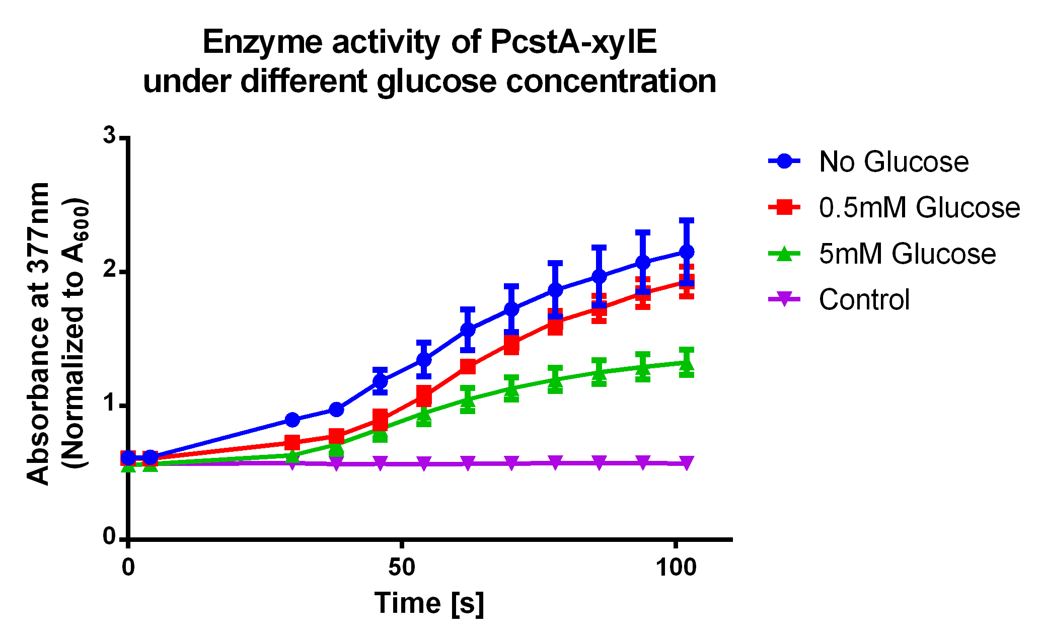
Before using xylE in this part for our project, we tested enzyme activity of this original part. We directly transformed the part plasmid into E. coli TOP10 strain, spread them on chloramphenicol plate, and picked single colony, which was then inoculated into 3ml chloramphenicol LB to culture overnight at 37℃.
Then the culture was separated into three parallel group and dilute to OD600 between 0.6-0.8, and different amount of glucose was added. After 1h of 220rpm shake at 37℃, the cell culture was aliquoted into 96-well plate, 100ul per well, 4 wells per group.
Varioscan Flash(TM) microplate reader was used to measure absorbance at 377nm of each well. The protocol for the instrument is listed here. The result showed significant decrease of group added 5 mM glucose than untreated group.
References
1Polissi A., Harayama S. (1993) In vivo reactivation of catechol 2,3-dioxygenase mediated by a chloroplast-type ferredoxin: a bacterial strategy to expand the substrate specificity of aromatic degradative pathways.. EMBO J., 12(8); 3339-3347
| None |

 1 Registry Star