Part:BBa_K3610045
EFR ectodomain / YFP
This part contains the ectodomain of the plant cell surface receptor EFR from A. thaliana fused to a yellow fluorescent protein. This part lacks the natural N-terminal signal sequence but instead uses the signal sequence from the alpha-Factor from yeast.
Contents
Usage and Biology
EFR
Elongation factor-thermo unstable receptor (EFR) from A. thaliana is a plant pattern-recognition receptor (PRR). It is a cell surface receptor and part of the plants first defence mechanism against potential pathogens. The EFR receptor is also a leucine-rich-repeats (LRR) receptor-like serine/threonine-protein kinase. The protein consists of an extracellular domain with leucine-rich repeats, a ligand binding domain found in many receptors, a single-pass transmembrane domain and finally an intracellular kinase domain. The ligand binding domain from EFR has high specificity to a bacterial pathogen-associated moleculat pattern (PAMP), namely the epitope elf18 of the abundant protein Elongation Factor Tu (EF-Tu), which is catalyzes the binding of aminoacyl-tRNA (aa-tRNA) to the ribosome in most prokaryotes and therefore is evolutionarily highly conserved. This makes the EFR a receptor that can be activated by the presence of a huge variety of bacteria. Upon binding of the ligand to the extracellular domain, the receptor dimerizes with its coreceptor BRI1-associated receptor kinase (BAK1). This interaction triggers the activation of the intracellular kinase domain of EFR and BAK1, initiating a signal cascade leading to an upregulation of immune response mechanisms.
EFR with YFP
In this sequence, the C-terminal domain entailing the intracellular kinase domain was replaced with the sequence coding for the yellow fluorescent protein venus, while the ectodomain and the transmembrane domain, including the juxtamembrane domain were kept. Additionally, a signal sequence native to S. cerevisiae was fused to the N-terminal sequence, which does not contain the native signal peptide. This way, the protein can be integrated into the membrane during translation and the expression can be observed as with the receptor protein, the YFP (Exλ : 515 nm, Emλ : 528 nm) gets translated as well.
Characterization
Expression of EFR ectodomain/YFP in S. cerevisiae
In a first step we inserted the single fragments making up this part into a plasmid with a gentamycin-3-acetyltransferase gene and transformed E. coli (DH10alpha) with the plasmids for amplification. In the next step we assembled the fragments in a plasmid with a spectinomycin acetyltransferase and amplified the plasmids again in the same E. coli strain. For this step we applied the techniques of Golden Gate Cloning to get the fragments in the right order into the plasmid. The restriction enzyme we chose was BsaI. For expressing this part consisting of YFP and the receptor protein, we initially intended to use promoters of different strength to get more quantitative data. Finally, we got the construct in a plasmid with a truncated version of the ADH1 promoter from S. cerevisiae. For termination, this part has the terminator sequence of the enolase 2 protein from S. cerevisiae. The plasmid also contained the TRP1 gene, which encodes phosphoribosylanthranilate isomerase, an enzyme that catalyzes the third step in tryptophan biosynthesis. This enabled us to use the same plasmid for expression in S. cerevisiae. We prepared a medium containing YNB and free amino acids, without tryptophan. S. cerevisiae cells (AP4) were transfected with the plasmid and then plated on the selective medium.
Microscopy
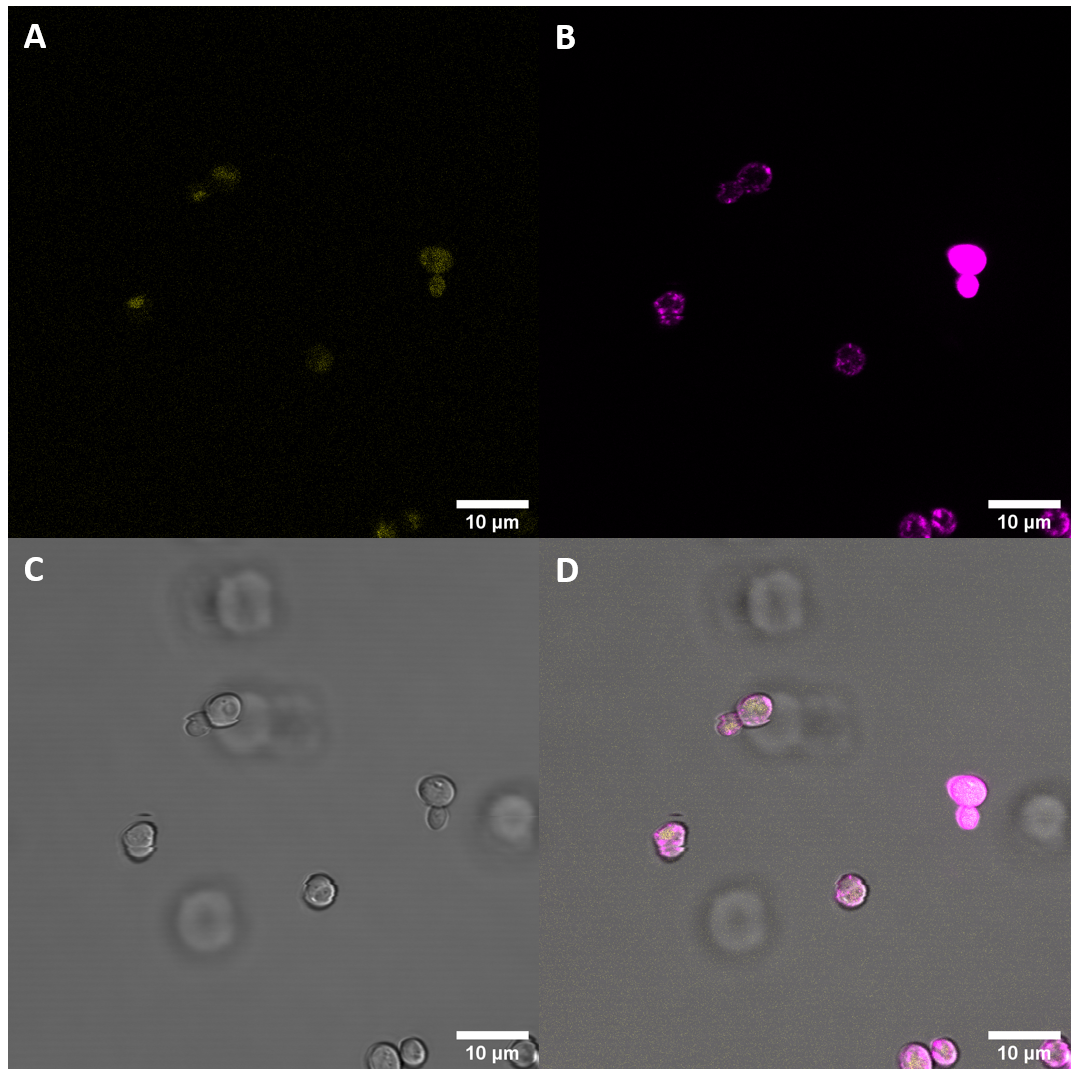
After successful transformation of yeast cells we checked for enhanced fluorescence with confocal fluorescence microscopy. If expression of YFP (λEx = 515 nm, λEx = 528 nm) can clearly be observed, it is reasonable to assume that the receptor domain is expressed as well, as the YFP is fused to the receptor protein. Expression of the construct was confirmed. We failed, however, to confirm localization at the cell membrane.
Imaging of the S. cerevisiae cells, which had been previously transfected with plasmids containing this construct, revealed increased fluorescence levels than the untransfected control. These results imply increased expression of YFP, indicating expression of the EFR ectodomain.
Additionally, the imaging results suggest that the proteins are in part localized at the cellular membrane, which is in alignment with our expectations as there is a secretion signal peptide and a transmembrane domain in the construct.
As localization at the cell membrane was something we were particularily interested in, we repeated the confocal microscopy step with an additional membrane stain. The cell membrane was stained with fm4-64, which fluoresces strongly after binding to the cell membrane ((λEX = 515nm and λEM = 640nm). The binding of the dye is happening rapidly and it is also reversible. If the time spent between staining and imaging is too long, then the dye will be taken up by the organism and stored in the vacuole. Imaging with a confocal microscope for YFP and the fm4-64 stain shows the spatial overlap of the red fluorescence of the stain and the yellow fluorescence of the protein fused to the receptors.
Confocal fluorescence microscopy with membrane staining confirmed what had been suggested previously with initial microscopy imaging. The results suggest that the receptor protein fused to the YFP gets epxpressed in S. cerevisiae and shows clear ring structures co-localized with the FM4-64 stain, indicating that there is localization at the plasma membrane.
Spectrometry
In addition to analyzing the cells with a microscope, we conducted a fluorescence assay with a plate reader. We conducted this experiment for multiple receptors at the same time. This way we were able to compare the expression levels of differnt versions of the BAK1 receptor. For each receptor we tried to isolate three different biological samples, however, not all cells grew. Ultimately, we only had two samples for the following S. cerevisiae cells: untransformed (Control), transformed with BAK1 ectodomain fused to YFP (eBAK) and the CORE ectodomain fused to YFP (eCORE). For the BAK1 with and without the native signal peptide fused to YFP (BAK+ and BAK-) and the EFR ectodomain fused to YFP (eEFR), we had samples from three different colonies. For each biological replicate, the optical density at absorbance of 600 nm (OD600) and the fluorescence levels were measured three times.
| measured OD600 values (OD) | |||||||||
|---|---|---|---|---|---|---|---|---|---|
| Replicate 1 | Replicate 2 | Replicate 3 | |||||||
| Blank | 0,08200000226 | 0,08200000226 | 0,08389999717 | ||||||
| Control | 0,3806000054 | 0,3747999966 | 0,4221999943 | 0,1316999942 | 0,131400004 | 0,1176000014 | |||
| BAK+ | 0,4943000078 | 0,4638999999 | 0,4514000118 | 0,5781000257 | 0,5253999829 | 0,5799999833 | 0,2615999877 | 0,2171999961 | 0,2011999935 |
| BAK- | 1,417099953 | 1,365499973 | 1,368899941 | 0,6305999756 | 0,5633999705 | 0,6216999888 | 0,896600008 | 0,7882999778 | 0,8032000065 |
| eBAK | 1,009699941 | 0,8404999971 | 0,8934999704 | 0,2653000057 | 0,2368000001 | 0,2592999935 | |||
| eCORE | 1,021499991 | 0,8616999984 | 0,9178000093 | 0,826300025 | 0,6888999939 | 0,7401999831 | |||
| eEFR | 1,379699945 | 1,322700024 | 1,333500028 | 1,035899997 | 1,014000058 | 0,9526000023 | 0,4860999882 | 0,3797000051 | 0,3829999864 |
The following settings were applied for fluorescence measurements:
| Mode: | Fluorescence Top Reading |
| Excitation Wavelength: | 485 nm |
| Emission Wavelength: | 535 nm |
| Excitation Bandwidth: | 20 nm |
| Emission Bandwidth: | 25 nm |
| Temperature: | 22.3°C |
| Fluorescence Top Reading (FTR) | |||||||||
|---|---|---|---|---|---|---|---|---|---|
| Replicate 1 | Replicate 2 | Replicate 3 | |||||||
| Blank | 1297 | 1282 | 1322 | ||||||
| Control | 2684 | 2474 | 2634 | 1852 | 1792 | 1750 | |||
| BAK+ | 3038 | 2813 | 2760 | 2836 | 2493 | 2788 | 2084 | 2072 | 2067 |
| BAK- | 35794 | 30319 | 31424 | 10792 | 9097 | 10517 | 22609 | 20227 | 21220 |
| eBAK | 26455 | 19828 | 21613 | 6614 | 5507 | 6229 | |||
| eCORE | 10709 | 8382 | 9339 | 8957 | 7062 | 7735 | |||
| eEFR | 43125 | 37782 | 39589 | 25641 | 24668 | 22517 | 12410 | 9054 | 9027 |
After measurement of the optical density and the fluorescence, the data were blank corrected (the average of the three blank measurements was subtracted from each measurement value).
The average of each of the three (or two) samples was calculated. From these values, the average was taken again.
After this step, we normalized the fluorescent output for OD600 (FTR/OD). The results of these calculations are displayed in the table below.
| Control | BAK+ | BAK- | eBAK | eCORE | eEFR |
|---|---|---|---|---|---|
| 4185,221063 | 9731,614266 | 26067,19254 | 28118,24739 | 3712,946478 | 23379,84399 |
If we set the values for the Control to 1 (Control = 1), then we get the fluorescence levels relative to the control, which is again diplayed in the table below.
| Control | eCORE | eEFR | BAK- | BAK+ | eBAK |
|---|---|---|---|---|---|
| 1 | 0,8871565975 | 5,586286516 | 6,228390841 | 2,325233033 | 6,718461693 |
Results of the plate reader suggested increased fluorescence through expression of YFP. The same was observed when cells were examined with the microscope. Therefore, S. cerevisiae cells express the plasmid containing the EFR ectodomain. Additionally, microscopy revealed localization at the cell membrane. This localization at the cell periphery is a big success as it shows, that the signal peptide from the alpha-Mating factor did, in fact induce translation into the membrane. Additionally, it was shown that the intracellular kinase domain is not necessary for membrane localization.
Flow Cytometry
It has been important to us to examine a sample with different approaches simultaneously, which is why we were eager to also measure fluorescence intensity by flow cytometry. In a first phase, 100,000 cells were measured from each biological replicate (488/530 FITC channel in a BD FACSCanto II flow cytometer). In the next phase, the biological replicates for each construct were pooled together and 200,000 cells from each sample were measured.
Flow catometry provided further evidence for expression of the construct in S. cerevisiae. Cells transfected with plasmids containing eEFR showed significantly increased fluorescence intensities when examined as single biological replicates, as well as when the replicates were pooled together in one sample.
During our project, expression of the ectodomain of EFR fused to YFP was confirmed with three different approaches. We used a fluorometric plate reader, which suggested a strong increase in fluorescence intesity and we also got similar results when measuring fluorescence with flow cytometry. In addition to these results, fluorescence imaging also showed increased fluorescence and about 25% of the cells showed a very clear ring structure which co-localized with the memrbane stain FM4-64. These results suggest that the receptor ectodomain can get expressed in S. cerevisiae and it also gets localized at the cell periphery, although a lot of the proteins get stuck and are localized within the cell.
These results are very promising and open many doors to future applications of this plant PRR in synthetic biology as they strongly suggest that expression of this receptor ectodomain and membrane localization (with the alpha-Factor signal peptide) can be achieved.
This expression of the EFR ectodomain in S. cerevisiae, which is accompanied by localization at the cell membrane is a big achievement and we hope that many future teams can profit from our insights.
Sequence and Features
- 10COMPATIBLE WITH RFC[10]
- 12INCOMPATIBLE WITH RFC[12]Illegal NheI site found at 355
Illegal NheI site found at 1285
Illegal NheI site found at 2203 - 21COMPATIBLE WITH RFC[21]
- 23COMPATIBLE WITH RFC[23]
- 25INCOMPATIBLE WITH RFC[25]Illegal NgoMIV site found at 1871
- 1000COMPATIBLE WITH RFC[1000]
| None |


