
File:FM.png
Revision as of 10:25, 9 October 2022 by Zeniazen5 (Talk | contribs) (Fluorescence microscope pictures of HEK 293T cells [A to F]. Scale is marked at the bottom left of the image. A: Negative control in Bright field B: Negative control overlapped with Bright field and DAPI staining, C: Positive control in GFP channel, D:...)
Size of this preview: 800 × 395 pixels. Other resolution: 320 × 158 pixels.
Original file (4,065 × 2,006 pixels, file size: 8.78 MB, MIME type: image/png)
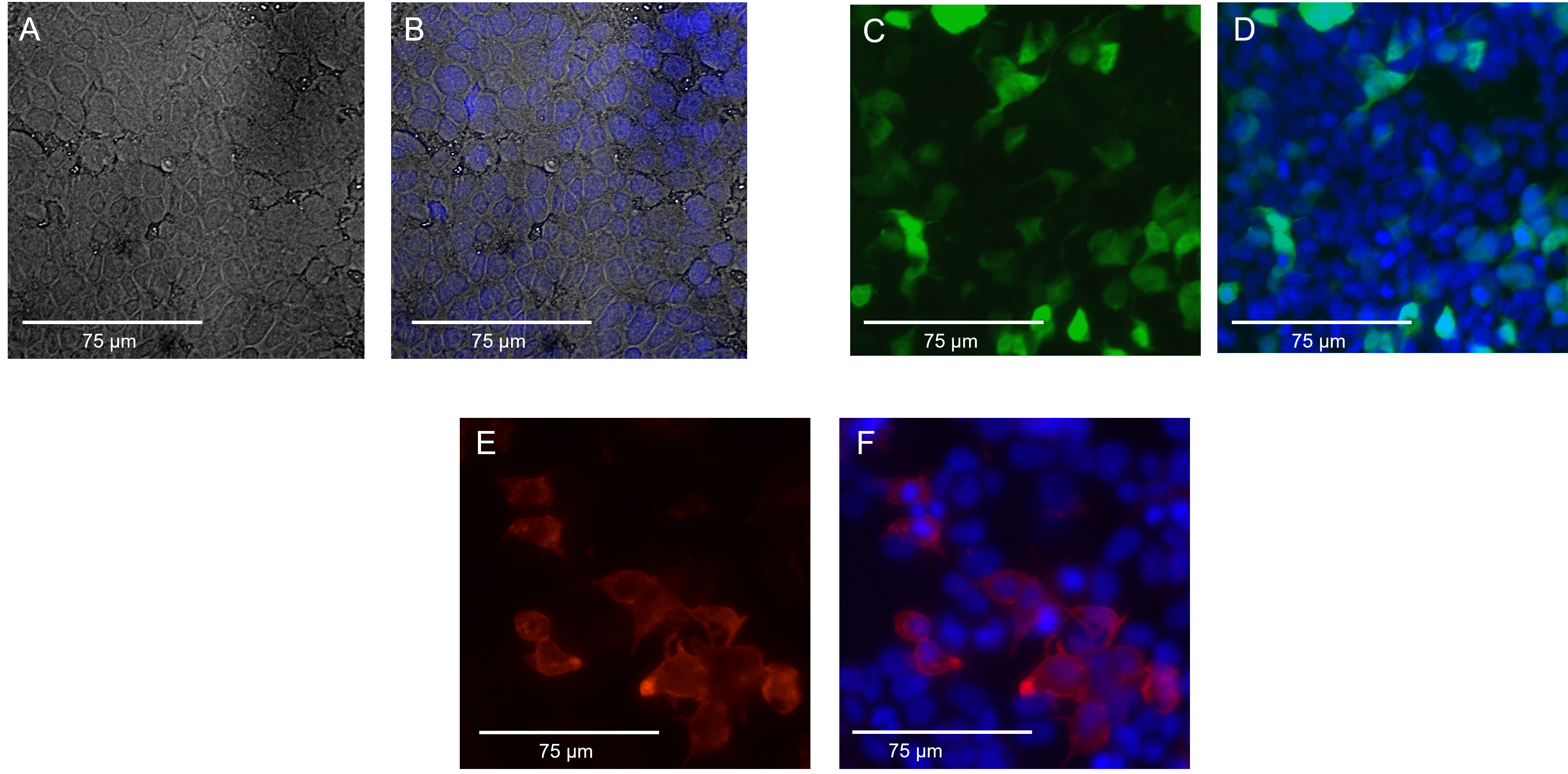
Fluorescence microscope pictures of HEK 293T cells [A to F]. Scale is marked at the bottom left of the image. A: Negative control in Bright field B: Negative control overlapped with Bright field and DAPI staining, C: Positive control in GFP channel, D: Positive control GFP superimposed with DAPI staining, E: Variant 20201 in RFP channel, F: Variant 20201 superimposed with IF and DAPI staining. The images were analyzed using FIJI-J imaging software [5].
File history
Click on a date/time to view the file as it appeared at that time.
| Date/Time | Thumbnail | Dimensions | User | Comment | |
|---|---|---|---|---|---|
| current | 10:25, 9 October 2022 | | 4,065 × 2,006 (8.78 MB) | Zeniazen5 (Talk | contribs) | Fluorescence microscope pictures of HEK 293T cells [A to F]. Scale is marked at the bottom left of the image. A: Negative control in Bright field B: Negative control overlapped with Bright field and DAPI staining, C: Positive control in GFP channel, D:... |
- You cannot overwrite this file.
File usage
The following page links to this file: