Part:BBa_K1592000
LIP2 prepro(signal peptide)
13 aa pre/4 XA or XP dipeptides/10 aa pro/KR cleavage site
The pre-region of lipase 2 from Yarrowia lipolytica corresponds to the signal sequence.
LIP2 prepro consist of 13aa pre-region of lipase2, four XA or XP dipeptides, 10aa pro-region of lipase2, and KR cleavage site.
the dipeptides XA and XP are substrates for diamino-peptidase; the dibasic KR cleavage site is substrate for Xpr6p endoproteinase.
Sequence and Features
- 10COMPATIBLE WITH RFC[10]
- 12COMPATIBLE WITH RFC[12]
- 21COMPATIBLE WITH RFC[21]
- 23COMPATIBLE WITH RFC[23]
- 25COMPATIBLE WITH RFC[25]
- 1000COMPATIBLE WITH RFC[1000]
Application of the part
This signal tag, LIP2 prepro, fused to Mcfp3 and constructed a new biobrick (BBa_K1592003), can lead the co-translational translocation of heterologous protein Mcfp-3 to be secreted out of the cell to achieve its function.
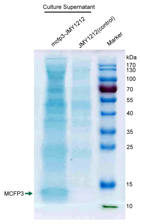
We concentrated cell culture solution of test Y.lipolytca JMY1212 cells transformed LIP2 prepro-Mcfp3 plasmid and control wildtype cells, and then separated proteins by SDS-PAGE.
Figure shows an obvious ~12kDa protein bands of LIP2 prepro-Mcfp3 in test lane, which cannot be found in control lane. This result proves that LIP2 prepro signal peptide can successfully lead the flocculating proteins Mcfp3 into surrounding environment.
Improvement
HUST-China 2015 put up a original method to cement sands as a promising way to help build firm structure in marine environment. The project “Euk.cement” was nominated “Best Environment Project” and “Best New Basic Part”. After the Jamboree, HUST-China iGEMers stepped forward--We did more part characterizations and achieved more valid data to submit to the registry. What’s more exciting is that we successfully published a paper “A living eukaryotic auto-cementation kit from surface display of silica binding peptides on Yarrowia lipolytica” on ACS Synthetic Biology(IF 6.076). We will demonstrate our working details in the following:
Flocculation system
Last year we ran a SDS-PAGE to identify the flocculating protein MCFP3. Moreover, we tested its function by applying the concentrated supernatant on an object slide and Microscopic observation after staining. The microscopy result qualitatively indicated the successfully secretion of MCFP3. This year, we made a further step-- BCA(bicinchoninic acid) quantification methods to describe the MCFP3 secretion level. We took wild Yarrowia lipolytica cultrue as control group to verify the proportion that MCFP3 accounted for in all secreted proteins. It was noteworthy that BCA quantification of the total protein in concentrated cell culture suspensions showed that the engineered cells released a protein level 0.1 µg/µL higher than that of the control group.
Sand cementation function
Last year, because of time limit we only tested sand cementation function of the ST123-JMY1212&mcfp3-JMY1212 mixed cells. It showed obvious effect on cementing sands. To make the data valid, this year, we together characterized 8 combinations of Si-tag domains and MCFP3 producing cells, and the result were quite corresponding to our expectation. The cementation test verified the cooperation of immobilization system and flocculation system cells in actual application conditions. Quartz sand (40 grams) mixed with either Si-tag+MCFP3 YMY1212 cells or control wild-type cells was loaded into a glass column, and a solution carrying oxygen, calcium and culture nutrients was supplied into the column using a peristaltic pump (Figure 4a). After 24 hours of treatment, the sand columns were dehydrated in a drying oven and then removed from the glass column. The sand treated with control wild-type JMY1212 cells was still scattered; only a few small clumps could be identified (Figure 4 c), and these may have been induced by the constitutive respiration of the wild type cells. However, with the treatment of Si-tag+MCFP3 cells, the sand aggregated, and an intact solid sand cylinder was obtained (Figure 4b). With further comparison of the treated sand under a microscope, the quartz sand granules treated with Si-tag+MCFP3 cells were found to be tightly agglutinated, whereas the quartz sand granules treated with wild type cells remained dispersed (Figure 4 d, e). This result indicated that Si-tag+MCFP3 cells actually worked well at making silica particles form a certain intact structure, which fits our hypothesis and design of their cementation function. It was also noticed in the cementation test that there were some small holes in the cemented sand cylinder. This special porous structure indicated the balance between the CO2 released from cell respiration and the calcium/magnesium sedimentation caused by the released CO2. This sedimentation, however, will be the final and vital step of the cementation process. Indeed, in some cementation applications, this structure is very important. For example, in desert sand consolidation treatment, this multi-porous structure will eliminate the potential compaction risk and will enable organisms to grow on it; in artificial reef construction on aqua farms, the multi-porous structure could also offer niches to all types of marine life.

To mimic the real conditions in underwater applications, we also tested the sand cementation under the condition of high water-to-sand ratio with turbulence in flasks on a shaker. Compared to sand treated with wild-type cells, sand treated with Si-tag cells and MCFP3 cells was found to be cemented together tightly using microscopy (Figure 4f, g). To find whether sand treated with different Si-tag cells can form cementation with different intensity, column cementation tests were also conducted with MCFP3 cells and different Si-tag cells. Sand treated with all Si-tag cells except wild-type control cells could form a cemented cylinder (Figure 4h). The relative intensity of the cylinders was quantified by the critical pressure value at cementation destruction. The results showed that Si-tag1+2+3 provided the highest cementation intensity, whereas Si-tag2+3 provided medium cementation intensity and the other strains provided weak cementation intensity (Figure 4i). This finding is comparable to the results from the Si-tag silica binding test in which Si-tag1+2+3 cells provided the highest silica binding intensity while other cells provided medium or weak intensity.
| None |


