Part:BBa_K1355001:Experience
This experience page is provided so that any user may enter their experience using this part.
Please enter
how you used this part and how it worked out.
Applications of BBa_K1355001
Experiments and Results as a Biosensor (BBa_K1355001 + BBa_E0840)
The experiment to quantify GFP expression induced by Hg was made according to the protocol “Quantification of Green Fluorescent Protein (GFP) induced by different concentrations of mercury in Escherichia coli DH5α”. DH5α transformed with BBa_K1355002 was inoculated in LM (LB with low concentration of NaCl) liquid medium with chloramphenicol and grew until the Optical Density was 0.4 to 0.6abs (measured on spectrophotometer at 600 nm wavelength). After cell growth, an aliquot of 500μl in 5 eppendorf tubes (2ml) was taken and then added mercury chloride in order to achieve the concentrations of: 0.01 µg/ml, 0.02 µg/ml, 0.1 µg/ml, 0.2 µg/ml, and 1 µg/ml. The samples were incubated at 37°C on shaker. We collected each eppendorf tube at time 1 (01:30 hours of incubation), time 2 (03:00 hours of incubation) and time 3 (04:30 hours of incubation). Every sample was centrifuged at 12000g for 3 minutes and the pellet washed with TN Buffer (Nacl 0.15M + Tris HCl 10mM) and then re-suspended with 500μl of the same buffer. The same process was made to the bacterium without construction as a control to GFP expression/intensity. GFP expression was measured using the Hidex Chameleon spectrofluorimeter with excitation filter 340 nm and emission filter 500 nm wavelength. The Optical Density was measured simultaneously. All samples were analyzed in triplicate.
The graph represented on Figure 1 shows the Optical Density of transformed DH5α with BBa_K1355002 in different Hg concentrations in function of time:
Figure 1. Optical Density measured in the four given times, at mercury chloride concentrations of 0 µg/ml, 0.01 µg/ml, 0.02 µg/ml, 0.1 µg/ml, 0.2 µg/ml, and 1 µg/ml.
In these condition cell growth increases along time. The highest values correspond to bacteria not exposed to mercury or to small concentrations, as in 0 µg/ml, 0.01 µg/ml and 0.02 µg/ml. Suggesting a harmless condition to bacteria. However, cell growth decreases at higher concentrations as, 0.1 µg/ml, 0.2 µg/ml and especially 1µg/ml, giving to bacteria a hard time for development.
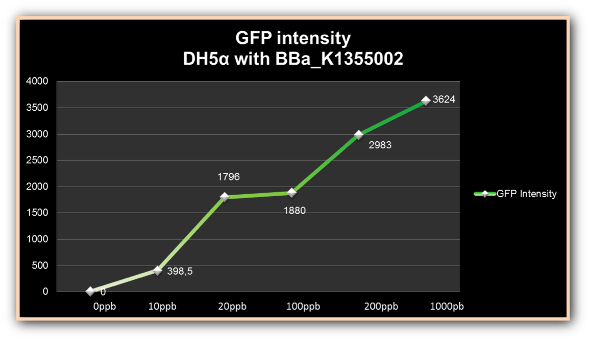
The graph represented on Figure 2 shows the fluorescence emitted by DH5-alpha induced by different Hg concentrations in function of the time; and the graph represented on Figure 3, shows the ratio between fluorescence emitted and Optical Density.
Figure 2. GFP fluorescence intensity in the four given times, at mercury chloride concentrations of 0 µg/ml, 0.01 µg/ml, 0.02 µg/ml, 0.1 µg/ml, 0.2 µg/ml, and 1 µg/ml.
Figure 3. GFP fluorescence per cell growth ratio in the four given times, at mercury chloride concentrations of 0 µg/ml, 0.01 µg/ml, 0.02 µg/ml, 0.1 µg/ml, 0.2 µg/ml, and 1 µg/ml.
Fluorescence can be observed in bacteria exposed to small concentrations, as 0.01µg/ml and 0.02µg/ml, but has low intensity. Fluorescence levels presents medium intensity in the higher concentration as consequence of cell death. Fluorescence intensity increased more than 480% in 0.2µg/ml concentrations, compared to fluorescence intensity from 1µg/ml, even in cell growth reduction, demonstrating its efficiency to induce mer promoter.
User Reviews
UNIQcef72f6198a6ced3-partinfo-00000000-QINU UNIQcef72f6198a6ced3-partinfo-00000001-QINU


