Part:BBa_K216005:Experience
This experience page is provided so that any user may enter their experience using this part.
Please enter
how you used this part and how it worked out.
Applications of BBa_K216005
Edinburgh iGEM team, October 2009: To test this part, it was combined with lacZ minigene J33202 for Miller assays (giving part BBa_K216009), and also with GFP for analysis using the promoter characterization kit of Jason Kelly. Results from overnight induction with Miller assay indicated stronger induction by nitrate than nitrite. A time course of induction using the GFP construct also indicated that nitrate gave stronger induction in the first few hours.
Results from Miller's Assay

Results from GFP promoter characterization kit
PyeaR response to nitrates
Jason Kelly’s assay demonstrates that our promoter is sensitive to nitrates. The most sensitive time period is between 5 ½ hours (320 minutes) 7 ½ hours (440 minutes) after induction. The sensitivity decreases with time, but induction is still at a high level after the peak activity of the promoter. Only two concentrations are shown (0 mM and 10 mM), as we have demonstrated that 10 mM is the sensitivity peak of this promoter (look at last graph).
PyeaR response to nitrites
Jason Kelly’s assay demonstrates that our promoter is sensitive to nitrites. The fluorescence levels begin to build up only after about 8 hours; therefore other irrelevant results are omitted from this graph. The sensitivity curve in this case is less steep compared to the one produced due to the presence of nitrates. This indicates that the promoter responds differently to these two nitrogenous compounds. Again, only two concentrations are shown (0 mM and 10 mM), as sensitivity peaks at 10mM.
PyeaR response to nitrates and nitrites after overnight induction
Here we can compare the activity of this promoter to different concentrations of both nitrates and nitrates. We observed that the promoter is activated more readily by nitrates compared to nitrites. Nevertheless, the activation produced by nitrites remains significant. The peak activation concentration for both compounds is 10 mM, after which activation efficiency decreases.

User Reviews
UNIQ9b22c4fc68d41ca8-partinfo-00000000-QINU UNIQ9b22c4fc68d41ca8-partinfo-00000001-QINU
NRP-UEA 2012
The team decided to develop the PYEAR biobrick further by ligating it to its mammalian counterpart: CArG promoter sequence E9-ns2. The genes were synthesised in two orientations, bacterial-mammalian (BBa_K774000) and mammalian-bacterial (BBa_K774001) as initially we were not sure what effect gene order would have on gene activity. The aim of this development was to increase the flexibility of the PYEAR promoter so that it can be used in both mammalian and bacterial systems. This is something that we thought was important as sensing nitric oxide in the human body has a wide range of therapeutic applications (please see the future applications section on our wiki).
GROWTH STUDIES
The two hybrid promoters were ligated with RFP (BBa_K774007 and BBa_K774005)and CFP (BBa_K774004 and BBa_K774006) in order to characterise them further.
This image (right) shows competent cells transformed with part: BBa K774004 and grown in media containing potassium nitrate (as a source of nitrates in order to induce promoter activity) at concentrations of 0 mM, 10 mM, 50 mM and 100 mM (from right to left). The E. coli was grown for 6 hours and then added to eppendorf tubes and spun down in a centrifuge in order to produce a pellet. The four samples were then viewed under a UV box to assess for fluorescence; as the photograph to the right shows that the sample at 0 mM potassium nitrate did not fluoresce, however those at 10, 50 and 100 mM potassium nitrate did fluoresce. They also appeared to fluoresce at the same strength, suggesting that 10 mM was equal to or above the maximum sensitivity level of this part.

The graph above shows the flourescence measured from the expression of eCFP due to the response of the bacterial-mammalian promoter to different concentrations of potassium nitrate. The wavelength reading which corresponds to eCFP is between 440-500nm. The graph clearly demonstrates that between 0mN and 15mM there is a proportional relationship between fluorescence intensity and potassium nitrate concentration. There appears to be a sharp increase in fluorescence intensity between 5mM and 10mM, and the rate at which intensity increase gradually decreases so that there is only a small increase between 15mM and 20mM.

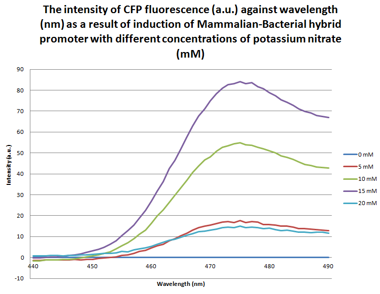
The graph above shows the flourescence measured from the expression of eCFP due to the response of the mammalian-bacterial promoter to different concentrations of potassium nitrate. The wavelength reading which corresponds to eCFP is between 440-500nm. The graph clearly demonstrates that between 0mN and 15mM there is a proportional relationship between fluorescence intensity and potassium nitrate concentration. It can be noted that at a 20mM concentration the intensity of fluorescence sharply decreases back down to the level of 5mM potassium nitate concentration. This may be due to the cell overexpressing eCFP up to the point at which the excess protein begins to form inclusion bodies which can no longer fluoresce; alternatively, this could be due the potassium nitrate concentration reaching the critical concentration at which it becomes toxic to the cell. This data differs to the readings taken from the bacterial-mammalian promoter ligated to eCFP, as well as the hybrid promoters to RFP, which may suggest there is a difference in the molecular mechanisms that these promoters function by; however at this point the change in intensity at 20mM is inconclusive and is an area which we would like to look into further.

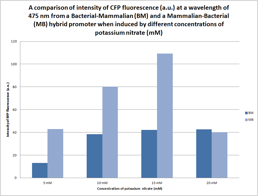
We were initially unsure of the effect that the orientation of the bacterial (pYEAR) and the mammalian (CaRG) genes would have in gene expression, therefore we synthesised two hybrid promoters in the orientation bacterial-mammalian and mammalian-bacterial. The graph above compares the intensity of fluorescence of the two hybrid promoters (BBa_K774004 and BBa_K774006) ligated to eCFP. There is a distinct difference between the intensity of fluorescence produced by the bacterial-mammalian promoter and the mammalian-promoter which is something that we would like to look into further. It is particularly interesting that at an intensity of 109a.u. the mammalian-bacterial promoter returns to the same level of intensity as the apparent maxiumum of the bacterial-mammalian promoter at 40a.u.

