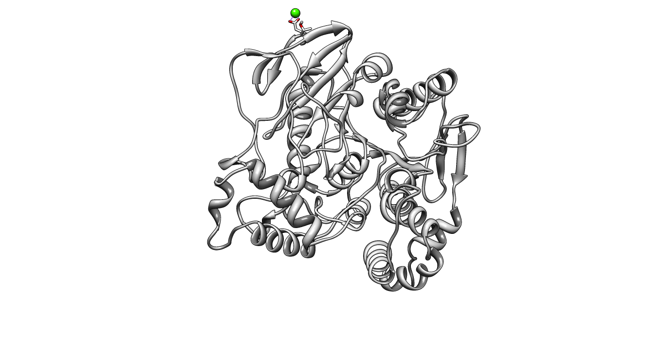
Part:BBa_K808026
pNB-Est 13: Esterase PET cleaving enzyme
The pNB-ESterase13 is a mutant derived from pNB-Esterase from Bacillus licheniformis. It showed the highest affinity and activity towards PET.
Usage and Biology
The characterization of pNB-Esterase13 (pNB-Est13) occured in 3 different ways.
Our [http://2012.igem.org/Team:TU_Darmstadt/Labjournal/Simulation Simulation Lab] performed
- [http://2012.igem.org/Team:TU_Darmstadt/Modeling_Docking#Docking Docking Simulations]
Our [http://2012.igem.org/Team:TU_Darmstadt/Project/Material_Science Material Science Lab] performed
- [http://2012.igem.org/Team:TU_Darmstadt/Labjournal/Material_Science#Surface_analysis_of_polyethylene_terephthalate_with_atomic_force_microscopy_.28AFM.29 Surface analysis of polyethylene terephthalat with atomic force microscopy]
Our [http://2012.igem.org/Team:TU_Darmstadt/Project/Degradation Degradation Lab]
- [http://2012.igem.org/Team:TU_Darmstadt/Labjournal/Degradation#Enzyme_Activity_Assays Enzyme activity assay]
Docking Simulation
Here, OET (dimeric state) became our reference substrate, meaning if an additive exhibits a higher inhibition constant it may effect our protein. Otherwise, if a binding energy exhibits a higher level than OET we can conclude that an additive may accumulate on our protein surface. Moreover, we compare the binding as well as the inhibition energies of the additives with itself to identify the strongest binder. Since we know the active site of the Fs. Cutinase and the PnB Est. 13, it will be interesting if a plastizicer may dock there. In order to analyze that, we counted the residues which are involved within every docking simulation. Therefore, we measured the distance of every ligand to its corresponding receptor with an appropriate threshold and thus identified the residues.
For the sake of convenience of this page, please visit our [http://2012.igem.org/Team:TU_Darmstadt/Modeling_Docking#Docking Simulation Lab > Docking Simulation] for further informtion.
Enzyme Activity Assay
pNP-Assay
We used a [http://2012.igem.org/Team:TU_Darmstadt/Protocols/pNP_Assay#pNP-Assay pNP-Assay] in order to describe.
Conditions were set at: T = 34°C and pH = 7.4. The method is simple. The absorption is measured every minute over 30 minutes by an ELISA reader capable of 96 well plates, after addition of enzyme. The released pNP-group has a typical absorption at 405nm wavelength.
Km and Vmax calculation
To find Km the growth of the first twelve data points were written down in another diagram. The slope of the regression line gave the speed of the hydrolysis for the single substrate concentrations. Now the value of the speed with the associated concentration of substrate were plotted in a Lineweaver-Burk diagram. At the point where the regression line is crossing the x-axis the x-coordinate amounts to -1/Km. So Km is about 400µM. To calculate the value of Vmax we used the point where the plotted line crosses the y-axis. The associated y-coordinate is 1/Vmax. Consequently Vmax for 5nM FsC is 0,001467 1/s.
Kcat calculation
The Kcat value is calculated through dividing Vmax by the enzyme concentration of 5nM. Kcat counts 0,293395 1/mol*s.
Indicator Assay
For a more visual proof of enzymatic activity we used Monomethyl terephthalate as a substrate and FsC as a catalyst. We used [http://en.wikipedia.org/wiki/Bromothymol_blue bromthymol blue] as an indicator. The release of COOH groups, due to hydrolytic degradation by FsC results in a colometric change from green to blue. For a more detailed description please visit our [http://2012.igem.org/Team:TU_Darmstadt/Labjournal/Degradation#Degradation_of_monomethyl_terephthalate_over_time labjournal> Degradation of monomethyl terephthalate]
PET degradation
Principle and conditions

Conditions: Room temperature (~ 22°C) pH = 7.4
Procedure
Round about 1g of PET granulate is added in each two falcon tubes to between 15 and 20 mL water of pH = 7.4 and several drops of bromthymol blue. In one of these falcon tubes Est13 stock solution is added until a concentration of 2,5 µM is reached. For 15 days the solutions are incubated at room temperature. At nine specific days of these the enzymatic solutions and the negative sample are photographed, to control the shift of pH.
Results
In all photos the degradation with Est13 is in the left falcon tube.
The photos show a decrease of the pH-value over time in the sample containing Est13. The one without this enzyme remains unchanged. So it is concluded that Est13 degrades PET granulate. Sequence and Features
- 10COMPATIBLE WITH RFC[10]
- 12COMPATIBLE WITH RFC[12]
- 21INCOMPATIBLE WITH RFC[21]Illegal BglII site found at 1071
- 23COMPATIBLE WITH RFC[23]
- 25INCOMPATIBLE WITH RFC[25]Illegal AgeI site found at 1142
- 1000COMPATIBLE WITH RFC[1000]
//function/degradation
| protein |


