Difference between revisions of "Part:BBa K404107"
| Line 42: | Line 42: | ||
AAV-293 cells were transfected with all genes necessary for producing viral particles encapsidating two different rAAV genomes with and without beta-globin intron respectively. mVenus expression was determined by flow cytometry 24-hours post infection of HT1080 cells. The rAAV genomes missing the beta-globin intron showed a negligible difference in mVenus expression compared to viral genomes containing the beta-globin intron. Considering these results, we suggest using the beta-globin intron in dependence on the size of your transgene. | AAV-293 cells were transfected with all genes necessary for producing viral particles encapsidating two different rAAV genomes with and without beta-globin intron respectively. mVenus expression was determined by flow cytometry 24-hours post infection of HT1080 cells. The rAAV genomes missing the beta-globin intron showed a negligible difference in mVenus expression compared to viral genomes containing the beta-globin intron. Considering these results, we suggest using the beta-globin intron in dependence on the size of your transgene. | ||
</p> | </p> | ||
| − | <img src="https://static.igem.org/mediawiki/parts/8/83/Freiburg10_FACS_withbetaglobin.png" width=" | + | <table> |
| − | height="auto"/><br /> | + | <tr> (anfang der ersten Zeile) |
| + | <td><img src="https://static.igem.org/mediawiki/parts/8/83/Freiburg10_FACS_withbetaglobin.png" width="400" | ||
| + | height="auto"/></td> | ||
| + | <td> Zwe<img src="https://static.igem.org/mediawiki/parts/8/83/Freiburg10_FACS_withbetaglobin.png" width="400" | ||
| + | height="auto"/><br /></td> | ||
| + | </tr> Ende der ersten Reihe | ||
| + | <tr> (ende der ersten Zeile) | ||
| + | <tr> | ||
| + | <td>Figure 9: Flow cytometry analysis of vectorplasmids with and without beta-globin intron. A: Gating non transduced cells (control); subcellular debris and clumps can be distinguished from single cells by size, estimated forward scatter (FS Lin) and granularity, estimated side scatter (SS Lin) B: Non transduced cells applied against mVenus (Analytical gate was set such that 1% or fewer of negative control cells fell within the positive region (R5). C: Gating transduced cells (R2 ≙R14) (used plasmids for transfection: GOI: pSB1C3_lITR_CMV_mVenus_hGH_rITR (BBa_K404128), pHelper, pRC). D: Transduced cells plotted against mVenus, R10 comprised transduced cells, by detecting mVenus expression E: Overlay of non-transduced (red) and transduced (green) cells applied against mVenus </td> | ||
| + | <td>F: Gating non-transduced cells (control). G: Non-transduced cells applied against mVenus (R5).H: Gating transduced cells (R2 ≙R14) (used plasmids for transfection: GOI: reassembled pSB1C3_lITR_CMV_beta-globin_mVenus_hGH_rITR (BBa_K404119), pHelper, pRC). I: Transduced cells applied against mVenus, R10 comprised transduced cells, by detecting mVenus expression. J: Overlay of non-transduced (red) and transduced (green) cells applied against mVenus.</td> | ||
| + | </tr> | ||
| + | </table> | ||
| − | <img src="https://static.igem.org/mediawiki/parts/ | + | <table> |
| − | height="auto"/><br /> | + | <tr> |
| − | + | <td> <img src="https://static.igem.org/mediawiki/parts/a/af/Freiburg10_Diagram_betaglobin.png" width="500" | |
| − | < | + | height="auto"/> <br /></td> |
| − | + | </tr> | |
| + | <tr> | ||
| + | <td>Figure 10: Flow cytometry analysis of vectorplasmids with and without beta-globin intron. 48-hours post transfection, viral particles were harvested by freeze-thaw lysis and centrifugation followed by HT1080 transduction. YFP expression of vectorplasmids was determined by flow cytometry 24-hours post infection. The vectorplasmid missing the beta-globin intron showed a negligible difference in mVenus expression compared to viral plasmid containing the beta-globin intron</td> | ||
| + | </tr> | ||
| + | </table> | ||
</html> | </html> | ||
| − | |||
| − | |||
<span class='h3bb'>Sequence and Features</span> | <span class='h3bb'>Sequence and Features</span> | ||
<partinfo>BBa_K404107 SequenceAndFeatures</partinfo> | <partinfo>BBa_K404107 SequenceAndFeatures</partinfo> | ||
Revision as of 21:07, 25 October 2010
Beta-Globin-Intron
Usage and Biology
| beta-globin intron | |
|---|---|

| |
| BioBrick Nr. | BBa_K404107 |
| RFC standard | RFC 10 |
| Requirement | pSB1C3 |
| Source | pAAV_MCS |
| Submitted by | [http://2010.igem.org/Team:Freiburg_Bioware FreiGEM 2010] |
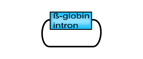
Providing an element assumed to be an enhancer of transgene expression (Nott, Meislin, & Moore, 2003), the iGEM team Freiburg presents a beta-globin intron derived from the human beta globin gene which can be fused upstream of the desired gene of interest.
Introns are non-coding sequences that are spliced after gene transcription. Apart from the possibility of alternative splicing which leads to an increased variability of translated proteins from one single gene, additional functions of introns have been found. They regulate and enhance gene expression at multiple levels such as initiating transcription, gene editing and polyadenylation of pre-mRNA (Nott, Le Hir, & Moore, 2004).In Valencia, Dias, & Reed (2008) the authors described the influence of splicing transgenes containing one intron at the 5´ and 3´ position, respectively. They demonstrated that splicing promotes the nuclear export of mRNA and that the spliceosome is cross-coupled to the mRNA export machinery.

Characterization
The BioBrick part beta-globin intron consists partially of a chimeric CMV promoter, followed by the intron II of the beta-globin gene. The 3´end of the intron is fused to the first 20 nucleotides of exon 3 of the beta globin gene. Our BioBrick part beta globin intron is assumed to enhance eukaryotic gene expression.
AAV-293 cells were transfected with all genes necessary for producing viral particles encapsidating two different rAAV genomes with and without beta-globin intron respectively. mVenus expression was determined by flow cytometry 24-hours post infection of HT1080 cells. The rAAV genomes missing the beta-globin intron showed a negligible difference in mVenus expression compared to viral genomes containing the beta-globin intron. Considering these results, we suggest using the beta-globin intron in dependence on the size of your transgene.
 |
Zwe |
| Figure 9: Flow cytometry analysis of vectorplasmids with and without beta-globin intron. A: Gating non transduced cells (control); subcellular debris and clumps can be distinguished from single cells by size, estimated forward scatter (FS Lin) and granularity, estimated side scatter (SS Lin) B: Non transduced cells applied against mVenus (Analytical gate was set such that 1% or fewer of negative control cells fell within the positive region (R5). C: Gating transduced cells (R2 ≙R14) (used plasmids for transfection: GOI: pSB1C3_lITR_CMV_mVenus_hGH_rITR (BBa_K404128), pHelper, pRC). D: Transduced cells plotted against mVenus, R10 comprised transduced cells, by detecting mVenus expression E: Overlay of non-transduced (red) and transduced (green) cells applied against mVenus | F: Gating non-transduced cells (control). G: Non-transduced cells applied against mVenus (R5).H: Gating transduced cells (R2 ≙R14) (used plasmids for transfection: GOI: reassembled pSB1C3_lITR_CMV_beta-globin_mVenus_hGH_rITR (BBa_K404119), pHelper, pRC). I: Transduced cells applied against mVenus, R10 comprised transduced cells, by detecting mVenus expression. J: Overlay of non-transduced (red) and transduced (green) cells applied against mVenus. |
 |
| Figure 10: Flow cytometry analysis of vectorplasmids with and without beta-globin intron. 48-hours post transfection, viral particles were harvested by freeze-thaw lysis and centrifugation followed by HT1080 transduction. YFP expression of vectorplasmids was determined by flow cytometry 24-hours post infection. The vectorplasmid missing the beta-globin intron showed a negligible difference in mVenus expression compared to viral plasmid containing the beta-globin intron |
- 10COMPATIBLE WITH RFC[10]
- 12COMPATIBLE WITH RFC[12]
- 21COMPATIBLE WITH RFC[21]
- 23COMPATIBLE WITH RFC[23]
- 25COMPATIBLE WITH RFC[25]
- 1000COMPATIBLE WITH RFC[1000]
References
Nott, A., Le Hir, H., & Moore, M. J. (2004). Splicing enhances translation in mammalian cells: an additional function of the exon junction complex. Genes & development, 18(2), 210-22. doi: 10.1101/gad.1163204.
Nott, A., Meislin, S. H., & Moore, M. J. (2003). A quantitative analysis of intron effects on mammalian gene expression. RNA (New York, N.Y.), 9(5), 607-17. doi: 10.1261/rna.5250403.et.
Valencia, P., Dias, A. P., & Reed, R. (2008). Splicing promotes rapid and efficient mRNA export in mammalian cells. Proceedings of the National Academy of Sciences of the United States of America, 105(9), 3386-91. doi: 10.1073/pnas.0800250105.
