Difference between revisions of "Part:BBa K525121"
(→Identification and localisation) |
|||
| Line 48: | Line 48: | ||
===Identification and localisation=== | ===Identification and localisation=== | ||
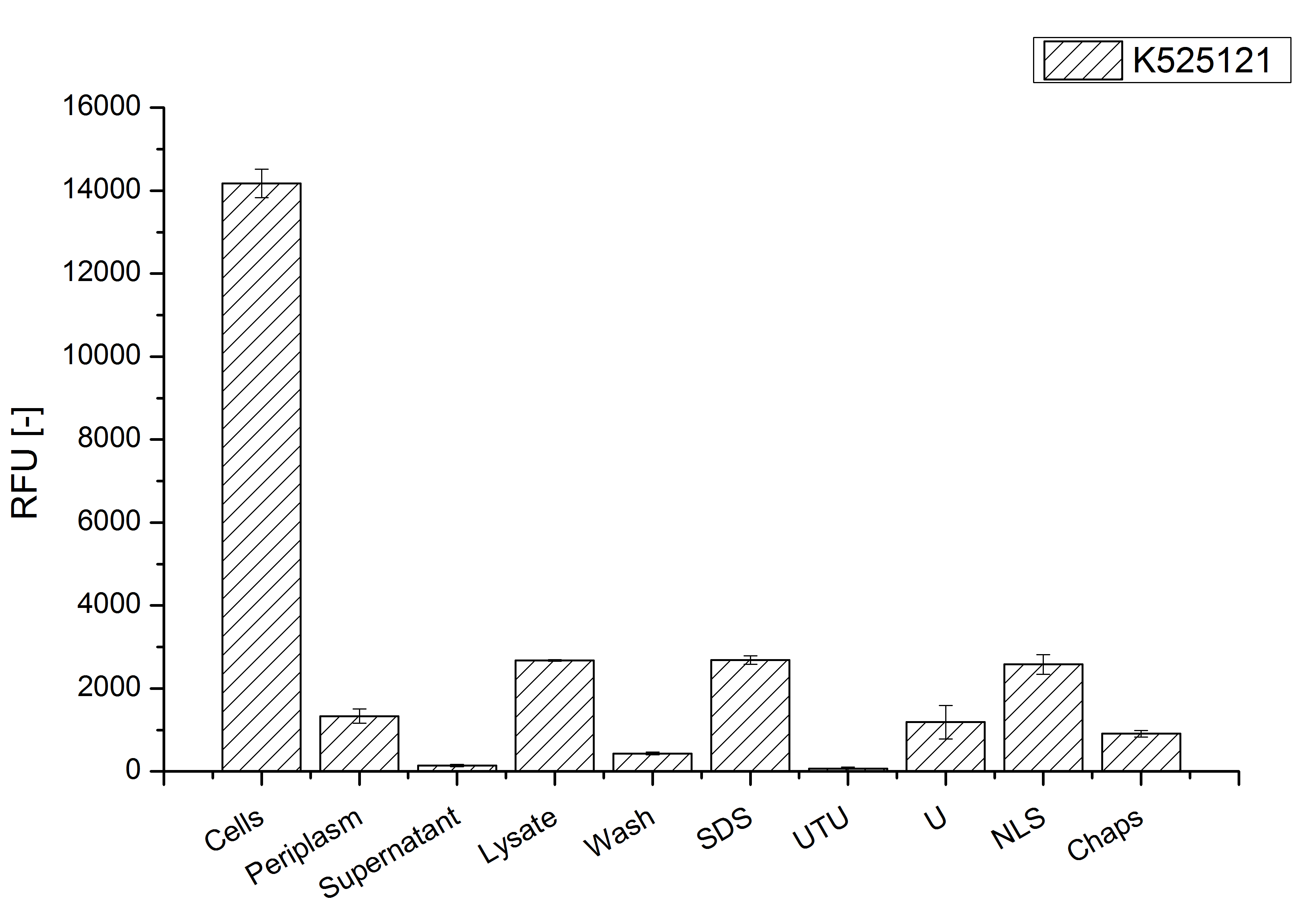
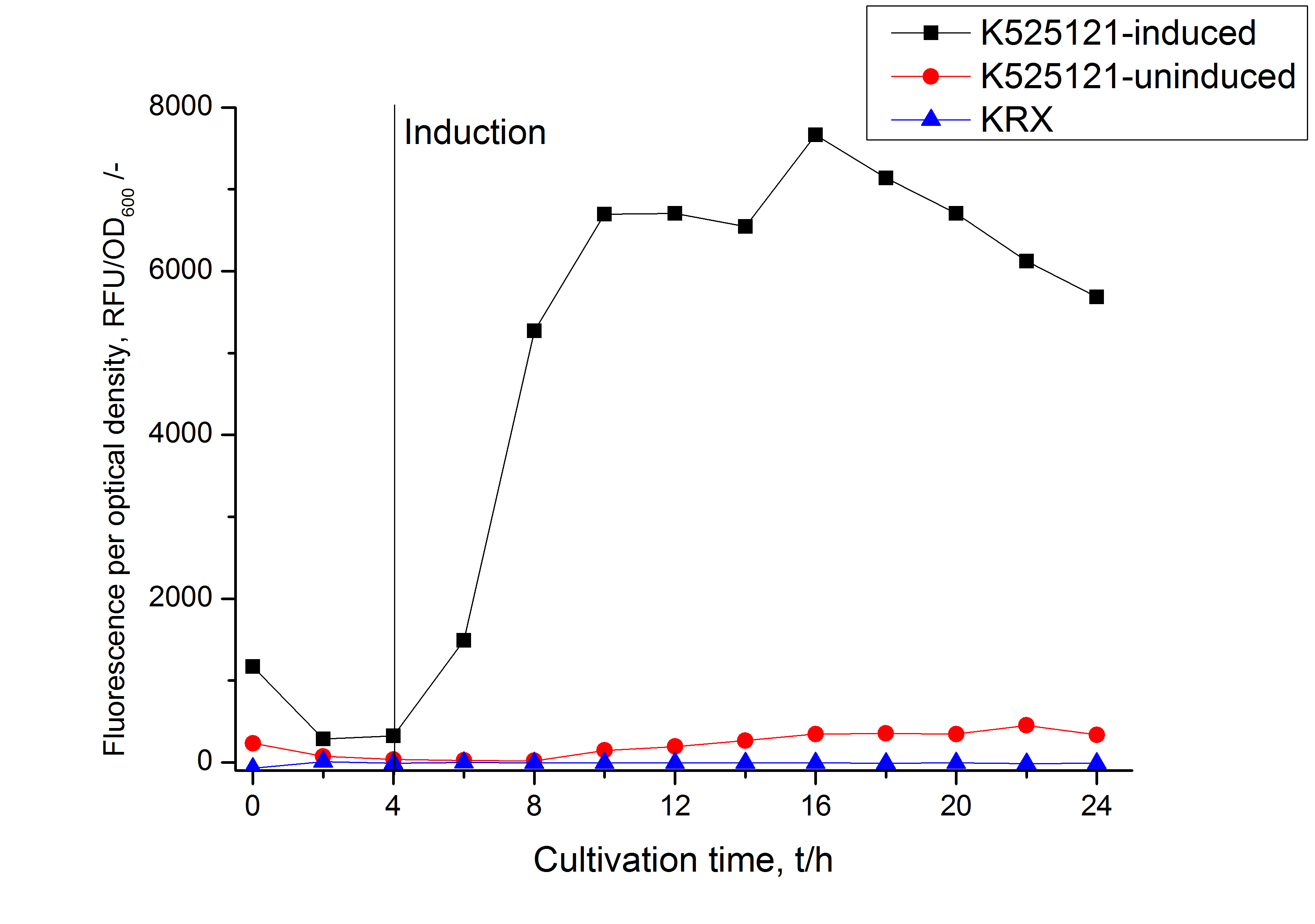
| − | After cultivation the CspB|mRFP fusion protein has to be localized in ''E. coli'' KRX. Therefor a part of the produced biomass was mechanically disrupted and the resulting lysat was wahed with ddH<sub>2</sub>O. From the other part the periplasm was isolated using a osmotic shock. The existance of fluorescene in the periplasma fraction, shown in fig. | + | After cultivation the CspB|mRFP fusion protein has to be localized in ''E. coli'' KRX. Therefor a part of the produced biomass was mechanically disrupted and the resulting lysat was wahed with ddH<sub>2</sub>O. From the other part the periplasm was isolated using a osmotic shock. The existance of fluorescene in the periplasma fraction, shown in fig. 3, indicate that ''Brevibacterium flavum''-signal sequence is at least in part functional in ''E. coli'' KRX. |
The S-layer fusion protein could not be found in the polyacrylamide gel after a [http://2011.igem.org/Team:Bielefeld-Germany/Protocols/Analytics#Sodium_dodecyl_sulfate_polyacrylamide_gel_electrophoresis_.28SDS-PAGE.29 SDS-PAGE] of the lysate and the cell depris were still red. This indicated that the fusion protein intigrates because of the lipid anchor into the cell membrane. For testing this assumption the washed lysate was treted with ionic, nonionic and zwitterionic detergents to release K525121 out of the membranes. | The S-layer fusion protein could not be found in the polyacrylamide gel after a [http://2011.igem.org/Team:Bielefeld-Germany/Protocols/Analytics#Sodium_dodecyl_sulfate_polyacrylamide_gel_electrophoresis_.28SDS-PAGE.29 SDS-PAGE] of the lysate and the cell depris were still red. This indicated that the fusion protein intigrates because of the lipid anchor into the cell membrane. For testing this assumption the washed lysate was treted with ionic, nonionic and zwitterionic detergents to release K525121 out of the membranes. | ||
| − | The proportionally high RFU in the detergent fractions and the MALDI-TOF analysis of the relevant size range in the polyacrylamid gel approved the insertion into the cell membrane (fig. | + | The proportionally high RFU in the detergent fractions and the MALDI-TOF analysis of the relevant size range in the polyacrylamid gel approved the insertion into the cell membrane (fig. 3). |
| − | [[Image:Bielefeld 2011 BF1 Purification.png|700px|thumb|center| '''Figure | + | [[Image:Bielefeld 2011 BF1 Purification.png|700px|thumb|center| '''Figure 3: Fluorescence pro OD<sub>600</sub> progression of the CspB/mRFP [https://parts.igem.org/Part:BBa_E1010 (BBa_E1010)] fusion protein initiating with the cultivation fractions up to the detergent fractions of the seperate denaturations. Cultivations were carried out in autoinduction medium at 37 ˚C. The cells were mechanically disrupted and the resulting biomass was wahed with ddH<sub>2</sub>O and resuspendet in the respective detergent. The used detergent acronyms stand for: SDS = 10 % (v/v) sodium dodecyl sulfate; UTU = 7 M urea and 3 M thiourea; U = 10 M urea; NLS = 10 % (v/v) n-lauroyl sarcosine; CHAPS = 3-[(3-cholamidopropyl)dimethylammonio]-1-propanesulfonate.''']] |
<!-- --> | <!-- --> | ||
Revision as of 17:17, 21 September 2011
S-layer cspB from Corynebacterium glutamicum with TAT-Sequence and lipid anchor, PT7 and RBS
Usage and Biology
S-layer proteins can be used as scaffold for nanobiotechnological applications and devices by e.g. fusing the S-layer's self-assembly domain to other functional protein domains. It is possible to coat surfaces and liposomes with S-layers. A big advantage of S-layers: after expressing in E. coli and purification, the nanobiotechnological system is cell-free. This enhances the biological security of a device.
This fluorescent S-layer fusion protein is used to characterize purification methods and the S-layer's ability to self-assemble on surfaces.
Important parameters
| Experiment | Characteristic | Result |
|---|---|---|
| Expression (E. coli) | Localisation | cell membrane |
| Compatibility | E. coli KRX | |
| Inductor for expression | L-rhamnose for induction of T7 polymerase | |
| Characteristics | Molecular weight | |
| Theoretical pI |
Sequence and Features
- 10COMPATIBLE WITH RFC[10]
- 12COMPATIBLE WITH RFC[12]
- 21INCOMPATIBLE WITH RFC[21]Illegal BamHI site found at 1421
Illegal XhoI site found at 248
Illegal XhoI site found at 866 - 23COMPATIBLE WITH RFC[23]
- 25COMPATIBLE WITH RFC[25]
- 1000INCOMPATIBLE WITH RFC[1000]Illegal BsaI.rc site found at 1400
Illegal SapI site found at 647
Illegal SapI site found at 859
Illegal SapI site found at 1407
Expression in E. coli
The CspB gen was fused with a monomeric RFP (BBa_E1010) using [http://2011.igem.org/Team:Bielefeld-Germany/Protocols#Gibson_assembly Gibson assembly] for characterization.
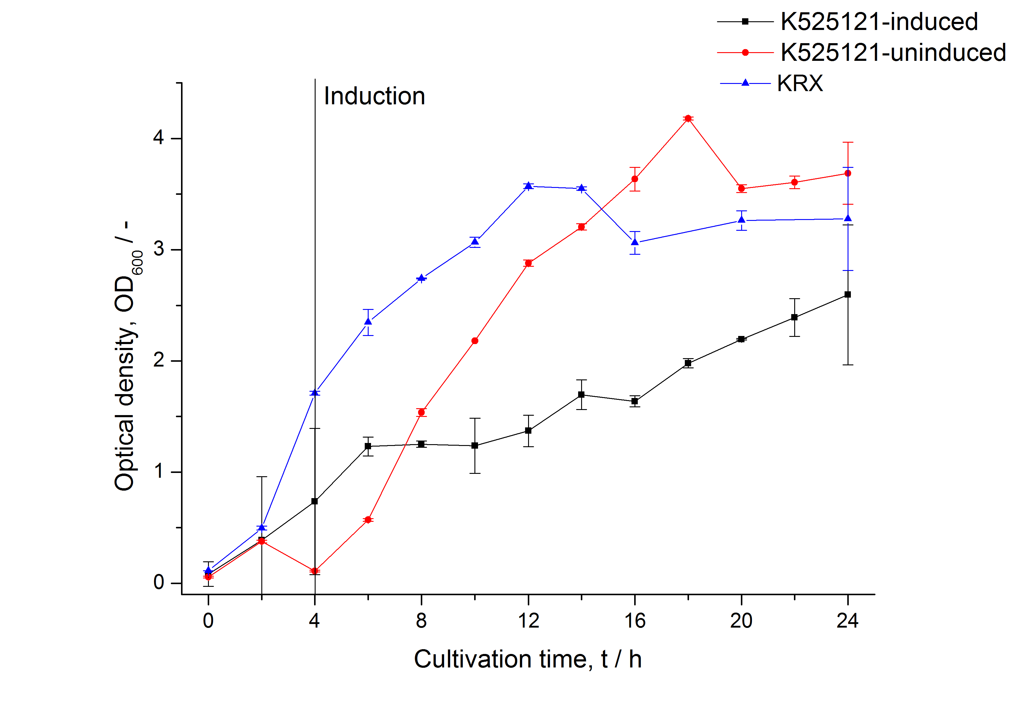
The CspB|mRFP fusion protein was overexpressed in E. coli KRX after induction of T7 polymerase by supplementation of 0,1 % L-rhamnose using the [http://2011.igem.org/Team:Bielefeld-Germany/Protocols/Downstream-processing#Expression_of_S-layer_genes_in_E._coli autinduction protocol] from promega.
Identification and localisation
After cultivation the CspB|mRFP fusion protein has to be localized in E. coli KRX. Therefor a part of the produced biomass was mechanically disrupted and the resulting lysat was wahed with ddH2O. From the other part the periplasm was isolated using a osmotic shock. The existance of fluorescene in the periplasma fraction, shown in fig. 3, indicate that Brevibacterium flavum-signal sequence is at least in part functional in E. coli KRX.
The S-layer fusion protein could not be found in the polyacrylamide gel after a [http://2011.igem.org/Team:Bielefeld-Germany/Protocols/Analytics#Sodium_dodecyl_sulfate_polyacrylamide_gel_electrophoresis_.28SDS-PAGE.29 SDS-PAGE] of the lysate and the cell depris were still red. This indicated that the fusion protein intigrates because of the lipid anchor into the cell membrane. For testing this assumption the washed lysate was treted with ionic, nonionic and zwitterionic detergents to release K525121 out of the membranes.
The proportionally high RFU in the detergent fractions and the MALDI-TOF analysis of the relevant size range in the polyacrylamid gel approved the insertion into the cell membrane (fig. 3).


