|
|
| Line 57: |
Line 57: |
| | ===Usage and Biology=== | | ===Usage and Biology=== |
| | | | |
| − | <!-- -->
| + | |
| | <span class='h3bb'>Sequence and Features</span> | | <span class='h3bb'>Sequence and Features</span> |
| | <partinfo>BBa_K2273036 SequenceAndFeatures</partinfo> | | <partinfo>BBa_K2273036 SequenceAndFeatures</partinfo> |
Revision as of 19:02, 1 November 2017
mCherry with C-Terminal SpyCatcher and His Tag
This composite part was used for evalutaion in the [http://2017.igem.org/Team:TU_Dresden/Project/Secretion secretion project] of 2017 TU_Dresden iGEM [http://2017.igem.org/Team:TU_Dresden team]. It codes for a fluorescent reporter protein (mCherry) and a functional tag (mini. SpyCatcher His-tagged), mediating covalent bonding with it tag partner (SpyTag). It is optimized for usage in Bacillus subtilis.
Design
A signal peptide (AmyE SP) was fused n-terminally to this construct to induce secretion.

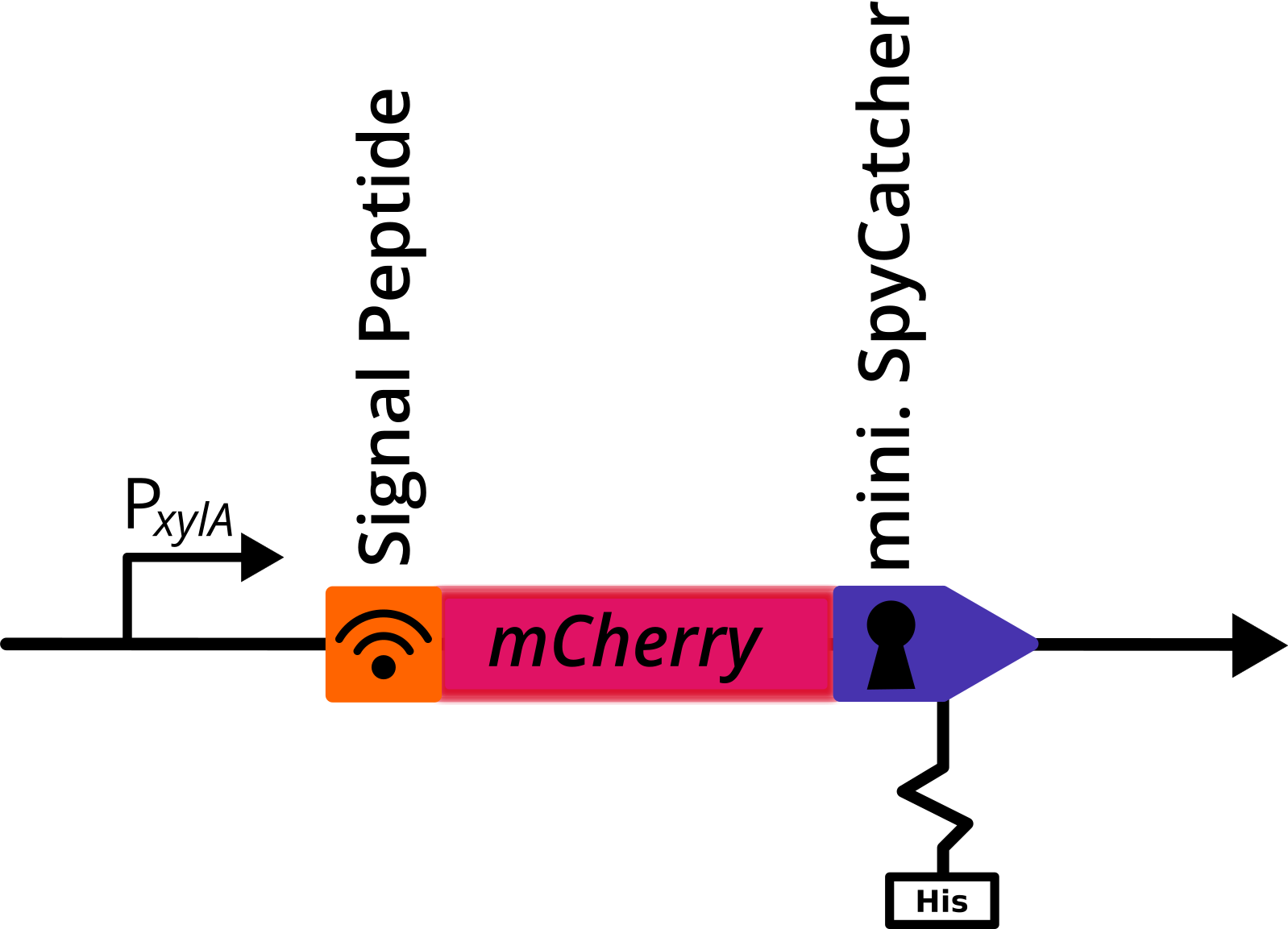
Figure 1:: Genetic construct with mCherry. Depicted is a translational fusion construct downstream of the
PxylA promotor, that was cloned in the multiple cloning site of the pBS2E
PxylA vector. The construct contains a signal peptide sequence, the gene coding for mCherry, c-terminally fused mini. SpyCatcher and a his-tag.
Application
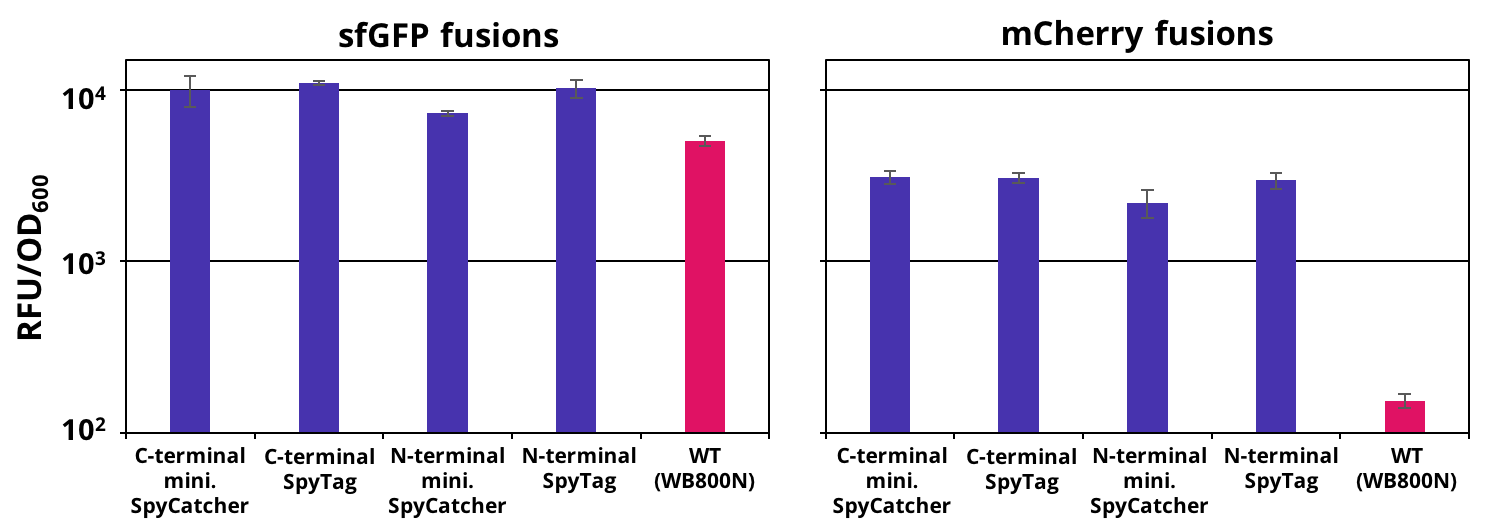
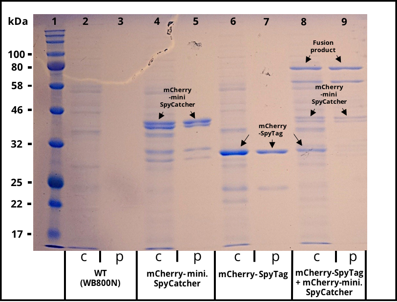
The successful secretion could be proven with a fluorescence assay using the supernatants of B. subtilis (Figure 2 and Figure 3). The functionality of the SpyTag/SpyCatcher was proven via SDS-PAGE, using the supernatants (Figure 4).

Figure 2:: Endpoint measurement of the fluorescence from supernatants carrying our constructs and the wild type. Expression of the single copy mCherry or sfGFP fusion SpyTag/SpyCather constructs (purple) was induced with 1% xylose and the supernatants were harvested after 16 h of incubation. Wild type supernatant is shown as a control (pink). Excitation wavelength for sfGFP was set to 480 nm and emission was recorded at 510 nm and for mCherry excitation wavelength was set to 585 nm and emission was recorded at 615 nm . The fluorescence was normalized by the optical density (OD600). Graph shows mean values and standard deviations of at least two biological and three technical replicates.

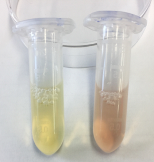
Figure 3:: Supernatants of B. subtilis cultures. Wild-type supernatant (left) and a mCherry-mini. SpyCatcher secreting strain (right). The expression of the multi-copy mCherry was induced with 1% Xylose and the supernatant was harvested after 16 h of incubation. .

Figure 4: SDS gel with crude and purified supernatants. Expression of the multi copy mCherry constructs was induced with 1% Xylose and the supernatants were harvested after 16 h of incubation. The his-tagged proteins were purified with Ni-NTA agarose beads. Lane 1 was loaded with 3 µl of NEB´s “Color Prestained Protein Standard Broad Range” ladder. Crude (c) and purified (p) supernatant of wild-type (WT) are shown as a control in lane 2 and 3. Lane 4 and 5 contain the supernatant of B. subtilis producing mCherry-mini. SpyCatcher fusion protein (36,6 kDa). Lane 4 and 5 contain the supernatant of B. subtilis producing mCherry-SpyTag fusion protein (31,9 kDa). The crude supernatants of the two mCherry producing strains were combined, incubated for 4 h, purified and loaded onto lane 8 and 9. The fusion product of the mCherry constructs is visable in the crude and purified supernatant.


