Difference between revisions of "Part:BBa K914018"
(→Characterization) |
|||
| Line 10: | Line 10: | ||
===Characterization=== | ===Characterization=== | ||
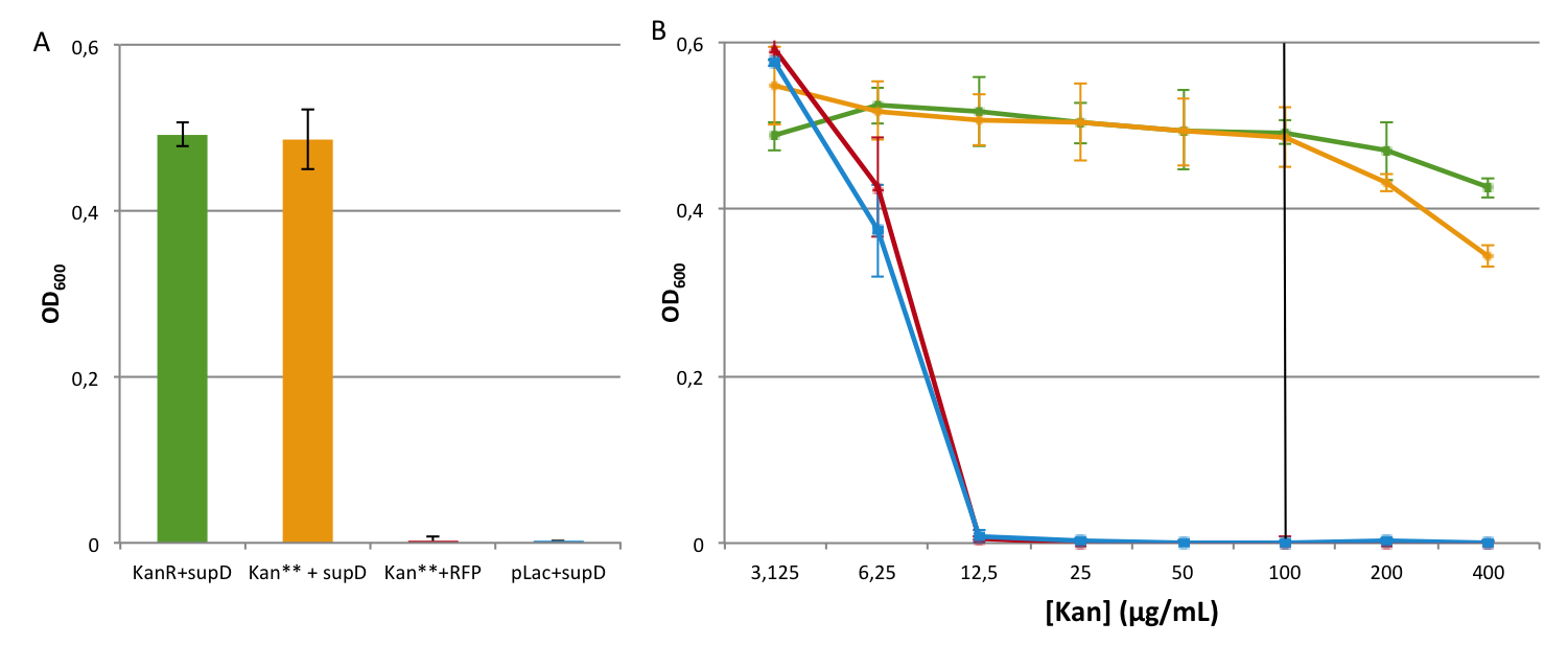
| + | The positive and negative controls behave correctly, and similarly to the previous experiment (see [https://parts.igem.org/Part:BBa_K914009:Experience K914009]), it is also the case for the complementation of Kan** with supD. That means that two mutation are not a problem for the cell. Moreover, we notice that there is no more leakiness in the system at usual concentration, even at low concentration. Actually the double kanamycin resistant gene with two mutation behave exactly like the negative control (Plac + supD) which means that semantic containment is working well. | ||
[[Image:SCquantitative**2.png|thumb|center|800px|'''Figure 4A and 4B:''' The bar graph represent the OD<sub>600</sub> at time = 8h20' (black line Figure 4C) for the different strains at a given kanamycin concentration (black line on Figure 4B). On B, we observe the variations of the OD<sub>600</sub> at different kanamycin concentrations, at the same time.]] | [[Image:SCquantitative**2.png|thumb|center|800px|'''Figure 4A and 4B:''' The bar graph represent the OD<sub>600</sub> at time = 8h20' (black line Figure 4C) for the different strains at a given kanamycin concentration (black line on Figure 4B). On B, we observe the variations of the OD<sub>600</sub> at different kanamycin concentrations, at the same time.]] | ||
| Line 15: | Line 16: | ||
[[Image:SCquantitative**1.png|thumb|center|800px|'''Figure 4C:''' The variations of the OD<sub>600</sub> is observed in function of the time, for different concentrations of kanamycin (400µg/mL, 100µg/mL, 25µg/mL).]] | [[Image:SCquantitative**1.png|thumb|center|800px|'''Figure 4C:''' The variations of the OD<sub>600</sub> is observed in function of the time, for different concentrations of kanamycin (400µg/mL, 100µg/mL, 25µg/mL).]] | ||
| − | + | ====Comparison of K914009 (Kan*) and K914018 (Kan**)==== | |
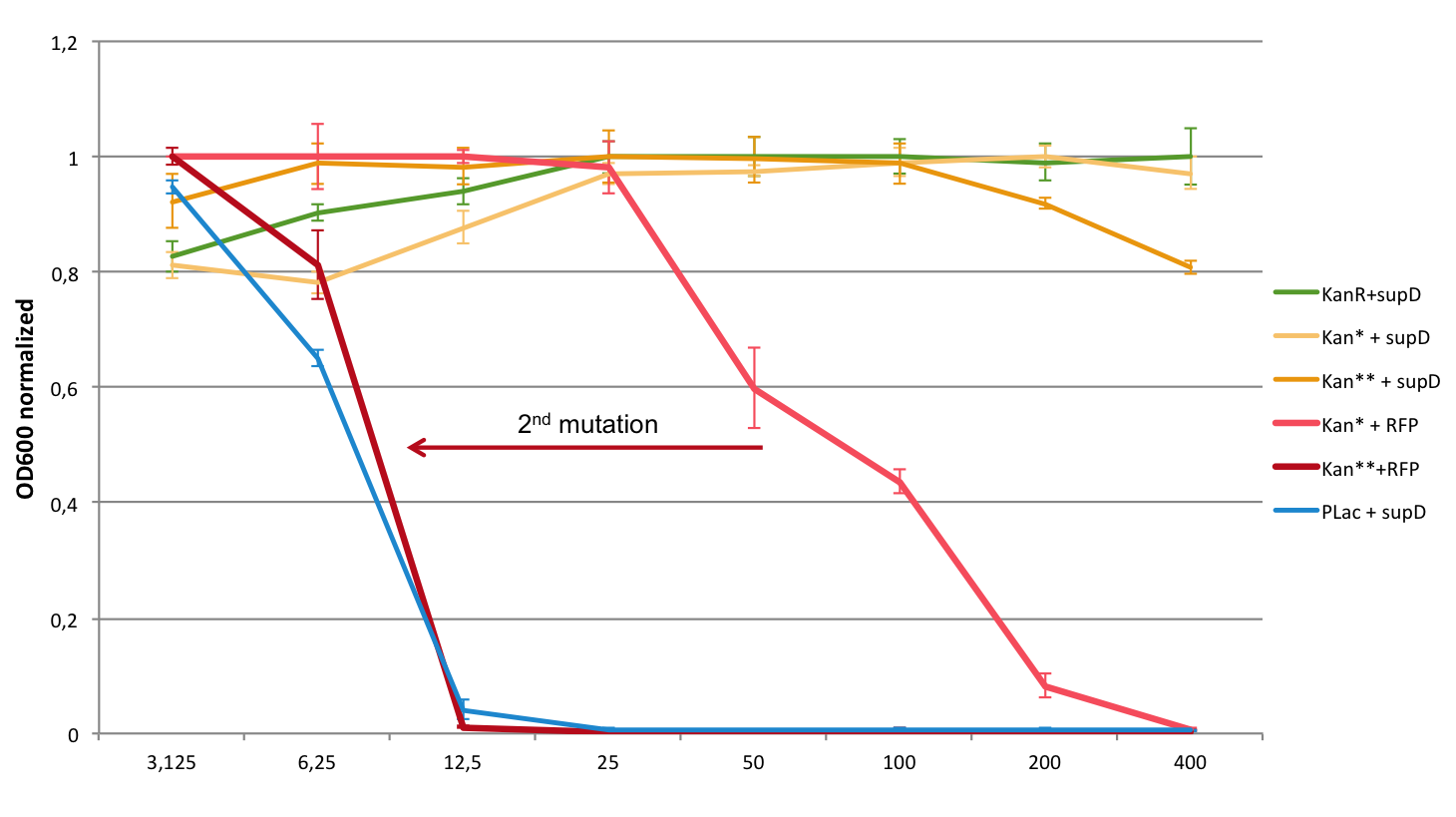
| + | Here is a graph that gather both results, to highlight the effect of the second mutation. The results are normalized intra group (experiment with the one-mutated gene, and with the two-mutated gene) in order to have the same scale to compare them. We took the average of the positive (KanR+supD) and the negative (Plac+supD) controls. | ||
| + | [[Image:SCquantitative**bilan.png|thumb|center|800px|'''Figure:''' Variation of OD<sub>600</sub> in different concentrations of [Kan] (µg/mL) at t= 8,37h]] | ||
<!--Uncomment this to enable Functional Parameter display --> | <!--Uncomment this to enable Functional Parameter display --> | ||
| + | |||
===Functional Parameters=== | ===Functional Parameters=== | ||
<partinfo>BBa_K914018 parameters</partinfo> | <partinfo>BBa_K914018 parameters</partinfo> | ||
<!-- --> | <!-- --> | ||
Revision as of 17:18, 28 October 2012
P1003** Kan resistant gene with 2 Amber Codon
The amber mutations avoid the expression of the P1003 gene even at usual concentration that prevent the expression of kanamycin resistance, although one mutation (BBa_K914009) permits some leakage in translation for this gene. This mutation is rescued with the tRNA amber suppressor supD (BBa_K914000). The idea here is to prevent the kanamycin gene resistance to be expressed in case horizontal gene transfer.
Sequence and Features
- 10COMPATIBLE WITH RFC[10]
- 12COMPATIBLE WITH RFC[12]
- 21COMPATIBLE WITH RFC[21]
- 23COMPATIBLE WITH RFC[23]
- 25COMPATIBLE WITH RFC[25]
- 1000COMPATIBLE WITH RFC[1000]
Characterization
The positive and negative controls behave correctly, and similarly to the previous experiment (see K914009), it is also the case for the complementation of Kan** with supD. That means that two mutation are not a problem for the cell. Moreover, we notice that there is no more leakiness in the system at usual concentration, even at low concentration. Actually the double kanamycin resistant gene with two mutation behave exactly like the negative control (Plac + supD) which means that semantic containment is working well.
Comparison of K914009 (Kan*) and K914018 (Kan**)
Here is a graph that gather both results, to highlight the effect of the second mutation. The results are normalized intra group (experiment with the one-mutated gene, and with the two-mutated gene) in order to have the same scale to compare them. We took the average of the positive (KanR+supD) and the negative (Plac+supD) controls.


