
File:T—TU Eindhoven--Platen part improvement.png
Size of this preview: 353 × 599 pixels. Other resolution: 141 × 240 pixels.
Original file (2,491 × 4,229 pixels, file size: 2.56 MB, MIME type: image/png)
File history
Click on a date/time to view the file as it appeared at that time.
| Date/Time | Thumbnail | Dimensions | User | Comment | |
|---|---|---|---|---|---|
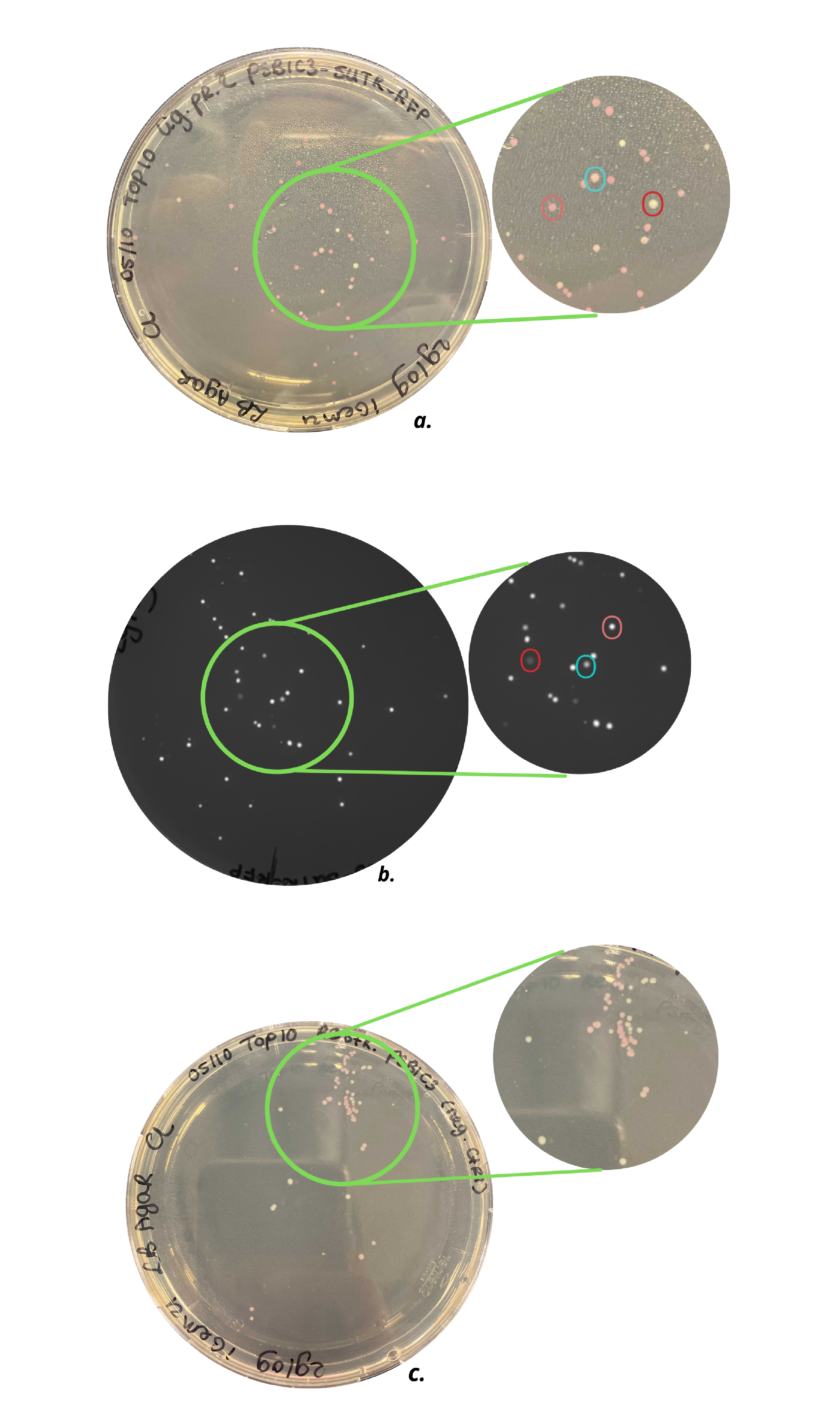
| current | 10:51, 19 October 2021 | | 2,491 × 4,229 (2.56 MB) | DaniekHoorn (Talk | contribs) | ‘’Figure 2. a) Top10 cells with pSB1C3-RFP plasmid, b) Top10 cells with pSB1C3-5’UTR-RFP plasmid under UV light, c) Top10 cells with a restricted pSB1C3-RFP plasmid (negative control). As can be seen in c) agar plate contains light pink and white... |
| 10:49, 19 October 2021 |  | 2,491 × 4,229 (2.56 MB) | DaniekHoorn (Talk | contribs) |
- You cannot overwrite this file.
File usage
The following 2 pages link to this file: