Difference between revisions of "Part:BBa K864402:Experience"
(→Applications of BBa_K864402) |
(→Applications of BBa_K864402) |
||
| (35 intermediate revisions by 5 users not shown) | |||
| Line 8: | Line 8: | ||
<h2> 2019 iGEM team Linkoping Sweden </h2> | <h2> 2019 iGEM team Linkoping Sweden </h2> | ||
| − | 2019 iGEM team Linkoping Sweden validated this part.<br><br> | + | 2019 iGEM team Linkoping Sweden validated this part.<br> |
| + | <html> | ||
| + | Summary: | ||
| + | In this contribution, we characterized the visual absorbance and the fluorescence of this construct. We also tested the oxygen dependency of the protein expression in <i>E. coli </i> BL21 (DE3) cells. The molecular weight of eforRed was also examined with an SDS-PAGE.<br> | ||
| + | <br> | ||
| + | |||
| + | <p> | ||
| + | <b style="font-size:120%;">Fluorescence and absorbance</b><br> | ||
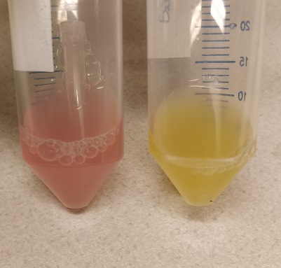
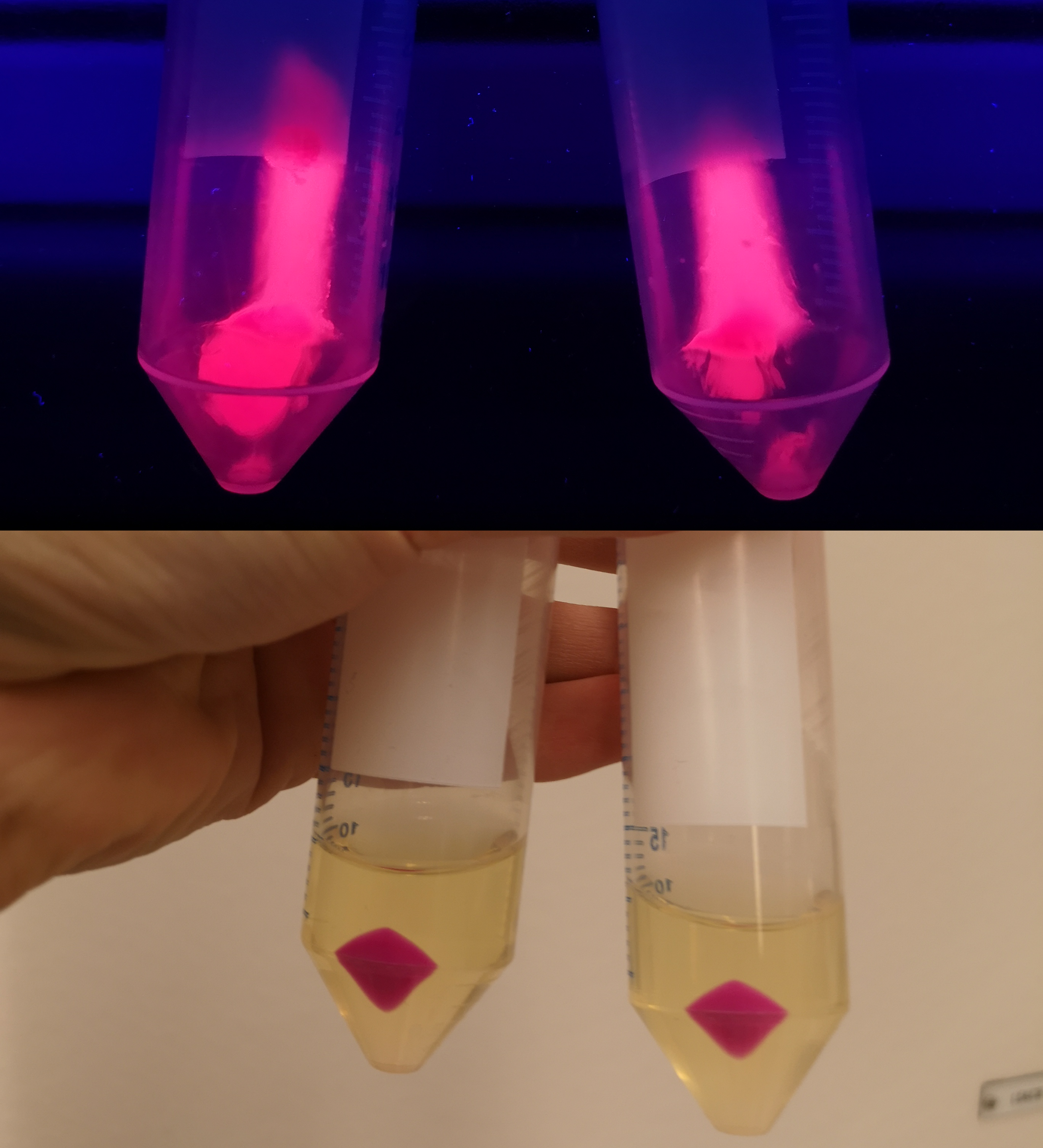
| + | To verify eforReds absorbance and emission, the construct was heat shocked into <i>Escherichia coli</i>, using the strain BL21 (DE3), and grown in a Falcon tube O.N. in 37 °C at 25 µg/mL chloramphenicol. Cotton plugs was used as corks for the Falcon tube. The bacterial solution was compared to a negative control with only BL21 (DE3)(Figure 1, left side) which showed the red color of eforRed in a bacterial solution. Thereafter, the eforRed expressing bacteria was centrifuged at 12 000 g for 10 minutes which displayed a burgundy colour (Figure 1, top-right corner). The pellet was also placed on an UV-table emitting light at 302 nm (Figure 1, down-right corner) and exhibited a pink glowing colour. These experiments verifies eforRed fluorescent effect as well as its absorbance in white light.</p> | ||
| − | |||
| − | |||
<br><br> | <br><br> | ||
| − | |||
| + | |||
| + | </html>[[Image:T--Linkoping_Sweden--pcons-eforred.jpeg|460px]]<html> | ||
| + | </html>[[Image:T--Linkoping_Sweden--eforred-pellet-photo.png|400px]]<html> | ||
<br><br> | <br><br> | ||
| − | + | <div class="figurtext" style=font-size:90%;><b>Figure 1.</b> The picture to the left depicts a cell culture of BL21 (DE3) pCons-eforRed (left tube) versus a negative control (right tube) with BL21 (DE3). The top right picture displays a pellet of BL21 (DE3) with pCons-eforRed in UV-light. The right picture is a pellet of BL21 (DE3) in white light.</div> | |
| − | + | </div> | |
| + | |||
<br><br> | <br><br> | ||
| − | |||
| + | Further characterization was performed in order to demonstrate the absorbance and fluorescence of eforRed. BL21 (DE3) containing pCons-eforRed were spread on a petri dish containing 25 µg/ml chloramphenicol and was photographed in white light and on an UV-table emitting 302 nm (Figure 2). The results were the same as above, in white light (Figure 2, right) the cultures had a burgundy color and on the UV-table the eforRed expressing bacteria exhibited a pink glowing colour (Figure 2, left). | ||
| − | + | <br><br> | |
| + | <div> | ||
| + | </html>[[Image:T--Linkoping_Sweden--eforred-agar-photo.png|700px|left|thumb|<b>Figure 2.</b> Colonies in the same host as previously is presented in white light (right side) and in 302 nm UV-light (left side).]]<html> | ||
| + | </div> | ||
| + | <br><br><br><br><br><br><br><br><br><br><br><br><br><br><br><br><br><br><br><br><br><br><br><br> | ||
| + | <div> | ||
| + | <p> | ||
| + | <b style="font-size:120%;">Oxygen dependency</b> | ||
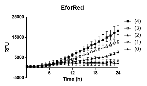
| + | To test the oxygen dependency of the protein production of eforRed in BL21 (DE3), the bacteria containing pCons-eforRed was grown O.N. to 2 OD<sub>600</sub> and diluted to 0.49 OD<sub>600</sub> with LB-miller. The bacteria was placed in a 96-well plate in replicates of 4 with 200 µL in each well. The oxygen access was varied by piercing different numbers of holes (0, 1, 2, 3 and 4) in the plastic film of the 96-well plate. A spectrometry experiment was conducted measuring the fluorescence (excitation 589, emission 609) in 37 °C for 24 hours and the experiment (Figure 3) showed that the access to oxygen effects the folding of eforRed and that 4 holes gave the highest yield.</p> | ||
| + | </div> | ||
| + | <br> | ||
| + | <div> | ||
| + | </html>[[Image:T--Linkoping_Sweden--efffforedd.png|460px]]<html> | ||
| + | </html>[[Image:T--Linkoping_Sweden--eforredvsmngphoto.jpeg|460px]]<html> | ||
| + | </div> | ||
| + | <br><br> | ||
| + | <div style=font-size:90%;> | ||
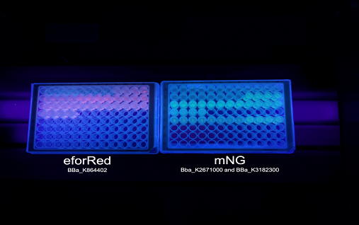
| + | <b>Figure 3.</b> To the left is a spectrometry experiment of BL21´s (DE3) protein production of eforRed with varying access to oxygen. The y-axis depicts the relative fluorescence intensity and the x-axis represents the time up to 24 hours. The plate with eforRed can be seen on the right pictures left side and the one on the right side is <i>E.coli</i> BL21 (DE3) with a different expression system used to test a strong green/yellow fluorescent protein. Both plates were illuminated in 302 nm UV-light. | ||
| + | </div> | ||
| − | <br><br><br><br> | + | <br><br> |
| + | <p><b style="font-size:120%;">Molecular weight</b><br> | ||
| + | A study of the molecular weight of pCons eforRed expressed in <i>E.coli</i> BL21 (DE3) was done by sonicating the cells and performing a SDS-PAGE electrophoresis on the lysate (<b>Figure 4.</b>). Biorads "Precision Plus Protein Dual Color Standards" was used as the protein ladder in the electrophoresis.<br><br> | ||
| + | </p> | ||
| + | </html>[[Image:T--Linkoping_Sweden--eforred_SDS-page.png|100px|left|]]<html> | ||
| + | <br><br><br><br><br><br><br><br><br><br><br><br><br><br><br><br><br> | ||
| + | <div class="figurtext"style=font-size:80%;> | ||
| + | <b>Figure 4.</b> SDS-page of sonicated <I>E.coli</I> BL21 (DE3) lysate with pCons-eforRed. Biorads "Precision Plus Protein Dual Color Standards" was used as the protein ladder. The visible band on the gel lies between 25 and 37 kD which corresponds to the molecular weight of eforRed which is 26.1 kDa. | ||
| + | </div> | ||
| + | </html> | ||
===User Reviews=== | ===User Reviews=== | ||
Latest revision as of 17:02, 20 October 2019
This experience page is provided so that any user may enter their experience using this part.
Please enter
how you used this part and how it worked out.
Applications of BBa_K864402
2019 iGEM team Linkoping Sweden
2019 iGEM team Linkoping Sweden validated this part.
Fluorescence and absorbance
To verify eforReds absorbance and emission, the construct was heat shocked into Escherichia coli, using the strain BL21 (DE3), and grown in a Falcon tube O.N. in 37 °C at 25 µg/mL chloramphenicol. Cotton plugs was used as corks for the Falcon tube. The bacterial solution was compared to a negative control with only BL21 (DE3)(Figure 1, left side) which showed the red color of eforRed in a bacterial solution. Thereafter, the eforRed expressing bacteria was centrifuged at 12 000 g for 10 minutes which displayed a burgundy colour (Figure 1, top-right corner). The pellet was also placed on an UV-table emitting light at 302 nm (Figure 1, down-right corner) and exhibited a pink glowing colour. These experiments verifies eforRed fluorescent effect as well as its absorbance in white light.


Further characterization was performed in order to demonstrate the absorbance and fluorescence of eforRed. BL21 (DE3) containing pCons-eforRed were spread on a petri dish containing 25 µg/ml chloramphenicol and was photographed in white light and on an UV-table emitting 302 nm (Figure 2). The results were the same as above, in white light (Figure 2, right) the cultures had a burgundy color and on the UV-table the eforRed expressing bacteria exhibited a pink glowing colour (Figure 2, left).
Oxygen dependency To test the oxygen dependency of the protein production of eforRed in BL21 (DE3), the bacteria containing pCons-eforRed was grown O.N. to 2 OD600 and diluted to 0.49 OD600 with LB-miller. The bacteria was placed in a 96-well plate in replicates of 4 with 200 µL in each well. The oxygen access was varied by piercing different numbers of holes (0, 1, 2, 3 and 4) in the plastic film of the 96-well plate. A spectrometry experiment was conducted measuring the fluorescence (excitation 589, emission 609) in 37 °C for 24 hours and the experiment (Figure 3) showed that the access to oxygen effects the folding of eforRed and that 4 holes gave the highest yield.
Molecular weight
A study of the molecular weight of pCons eforRed expressed in E.coli BL21 (DE3) was done by sonicating the cells and performing a SDS-PAGE electrophoresis on the lysate (Figure 4.). Biorads "Precision Plus Protein Dual Color Standards" was used as the protein ladder in the electrophoresis.
User Reviews
UNIQd2ab2d533c793235-partinfo-00000007-QINU UNIQd2ab2d533c793235-partinfo-00000008-QINU


