
Difference between revisions of "Part:BBa K1094401"
Emilfischer (Talk | contribs) |
m (Aachen 2019 edit) |
||
| Line 52: | Line 52: | ||
</html> | </html> | ||
| + | ===Usage and Biology=== | ||
<!-- --> | <!-- --> | ||
Revision as of 15:54, 20 October 2019
MamC-eGFP fusion
MamC (BBa_K1094001) from Magnetospirillum magnetotacticum (MS-1) tagged with enhanced green fluorescent protein (eGFP, BBa_K1094400). Between the parts a glycine linker (10 glycine residues) is added. The gene product can be used to detect localization of MamC in MS-1. A previous study on the related strain Magnetospirillum gryphiswaldense (MSR-1) showed the MSR-1 homologue of MamC to localize in the magnetosome membrane when fused to eGFP (Lang, C. & Schüler, D. "Expression of Green Fluorescent Protein Fused to Magnetosome Proteins in Microaerophilic Magnetotactic Bacteria". Applied and Environmental Microbiology. Aug. 2008, p. 4944-4953).
The two parts was cloned together using classical restriction digestion cloning in pBBR1MCS-2 in E. coli. The composite part was later PCR amplified and cloned into the expression vector pJAM1786 via pDONR207 in E. coli, by using the Gateway system by Invitrogen. Colony PCR and gel electrophoresis was performed to ensure the insert was in the vector.
Confocal microscopy was carried out. This gave rise to the pictures below. Left showing E. coli cells and right showing the fluorescence of these (defined in software to be red).


Eight colonies from the restreak from colony PCR were inoculated into liquid culture and grown overnight alongside three E. coli cultures containing a non-expression plasmid with a non-fluorescent insert (MamC-pSB1C3). Fluorescence was measured in an opaque ELISA reader plate. The following dilutions were made: 1X, 2X, 5X, 10X and 50X. OD(600nm) was measured to ensure similar cell density. Excitation was carried out at 485 nm and emission was collected at 535 nm.
The mean values of the fluorescence can be found in the table below. Standard error for both groups (Total SE) was assessed using a Satterthwaite approximation.

(The raw data can be found here)
All dilution showed significantly more fluorescence than the control cultures except for the 50X dilution. The mean of the difference to the control cultures is shown as a function of dilution below.

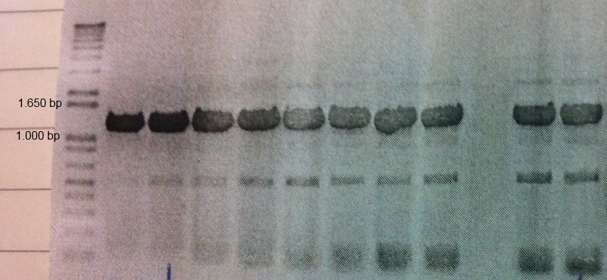
A Western blot of the MamC-eGFP-pJAM1786 cultures was made with anti-GFP as primary antibodies. The blot (shown below) gave rise to two bands. It is likely that MamC-eGFP is the larger band (approx. 40kDa). The smaller band was unexpected but can possibly be due to cleavage of the fusion protein.

Usage and Biology
Sequence and Features
- 10COMPATIBLE WITH RFC[10]
- 12COMPATIBLE WITH RFC[12]
- 21COMPATIBLE WITH RFC[21]
- 23COMPATIBLE WITH RFC[23]
- 25INCOMPATIBLE WITH RFC[25]Illegal NgoMIV site found at 169
Illegal NgoMIV site found at 253 - 1000COMPATIBLE WITH RFC[1000]