
Search results (Hint: Try the special 'Search Parts' system on the main page)
Create the page "Crystal" on this wiki! See also the search results found.
Page title matches

File:ChlP crystal structure.png (601 × 686 (497 KB)) - 01:02, 16 October 2014
File:Crystal Structure of Cyanobacterial ChlM.png (794 × 733 (396 KB)) - 05:57, 16 October 2014
File:ChlI protein crystal structure.png (133 × 133 (19 KB)) - 07:42, 17 October 2014
File:96 wells plate Crystal Violet K1583100.jpg 96 Wells plate of Crystal Violet Assay(723 × 518 (62 KB)) - 08:22, 18 September 2015
File:Crystal Violet result K1583100.JPG Graph showing the results of the crystal violet assay(1,181 × 550 (45 KB)) - 08:26, 18 September 2015
File:Crystal Violet his.png Crystal Violet assay of CsgA_His(1,069 × 761 (15 KB)) - 09:19, 18 September 2015
File:T--SCUT China--Crystal structure of archaeal toxin-antitoxin RelE-RelB complex .jpg (834 × 577 (38 KB)) - 08:11, 4 July 2020
File:Crystal structure of MerR (Rasmol).png (422 × 327 (70 KB)) - 18:48, 16 October 2020


.png)
Page text matches

File:TPcon1.jpg ...ze a temperature-regulated mechanism to control the expression period of a crystal protein. Therefore, we took advantage of the interaction between 37° C ind ...ate and growth rate, is highly dependent on amplification conditions, thus crystal protein production is off during the incubation/amplification step. Because(662 × 243 (22 KB)) - 05:06, 24 October 2010
File:C2001.jpg Crystal Structure of a Zif23-GCN4 Chimera Bound to DNA created by the Protein Datab(250 × 250 (11 KB)) - 23:57, 15 July 2006
File:1f39 bio r 250.jpg CRYSTAL STRUCTURE OF THE LAMBDA REPRESSOR C-TERMINAL DOMAIN as determined by Bell e(250 × 250 (12 KB)) - 20:32, 24 July 2006
File:Hin dimer closed 1GDT.gif Yang, W., Steitz, T.A. (1995) ''Crystal structure of the site-specific recombinase gamma delta resolvase complexed(549 × 547 (28 KB)) - 18:24, 29 October 2006
File:BCCS fhuA protein structure.jpg ...et al.(1998),Transmembranesignalling across the Ligand-Gated FhuAReceptor: Crystal structures of Free and Ferrichrome-Bound States reveal allostericchanges. C(575 × 510 (64 KB)) - 17:50, 14 July 2009
File:TECON F4.jpg ...We expect our circuit design to allow steady bacteria growth and inhibit crystal protein at T>37° C, and high protein production and low bacteria growth ra(560 × 408 (29 KB)) - 05:02, 24 October 2010
File:PCD Fischle 2003.tif Crystal structure of Drosophila melanogaster Pc Polycomb chromodomain. Figure 6 fro(202 × 192 (56 KB)) - 05:01, 3 July 2011
File:Peking R YXW Figure4.jpg ...Mary R. Stahley, Anne B. Kosek, Jimin Wang, and Scott A. Strobel. (2004). Crystal structure of a self-splicing group I intron with both exons. Nature 430, 45(829 × 614 (279 KB)) - 15:18, 5 October 2011
File:Esta autotransporter.png Van Den Berg, B. (2010). Crystal structure of a full-length autotransporter. Journal of Molecular Biology, 3(776 × 590 (313 KB)) - 15:20, 17 October 2011File:96 wells plate Crystal Violet K1583100.jpg 96 Wells plate of Crystal Violet Assay(723 × 518 (62 KB)) - 08:22, 18 September 2015File:Crystal Violet result K1583100.JPG Graph showing the results of the crystal violet assay(1,181 × 550 (45 KB)) - 08:26, 18 September 2015File:Crystal Violet his.png Crystal Violet assay of CsgA_His(1,069 × 761 (15 KB)) - 09:19, 18 September 2015
File:Nrfa protein structure IITD.png Crystal Structure of the Assimilatory Nitrite Reductase from RCSB-PDB.(226 × 248 (53 KB)) - 12:57, 18 September 2015
File:BBa K118003-Evry2016.jpeg Crystal structure of Phytoene desaturase CRTI from Pantoea ananatis(500 × 500 (39 KB)) - 23:06, 29 August 2016
File:T--Uppsala-4I3B.png Crystal structure of UnaG (1.2 Å resolution) with bound bilirubin. PDBid 4I3B.(607 × 700 (171 KB)) - 13:39, 18 October 2016
File:T--Goettingen--BBa K2586007 aroAstructure.png Crystal structure of Phospho-2-dehydro-3-deoxyheptonate aldolase (DAHP synthase) (T(500 × 500 (76 KB)) - 15:28, 23 September 2018
File:T--goettingen--BBa K2586007 structureAroE.jpg Crystal structure of putative 5-enolpyruvoylshikimate-3-phosphate synthase from Bac(500 × 500 (74 KB)) - 16:25, 23 September 2018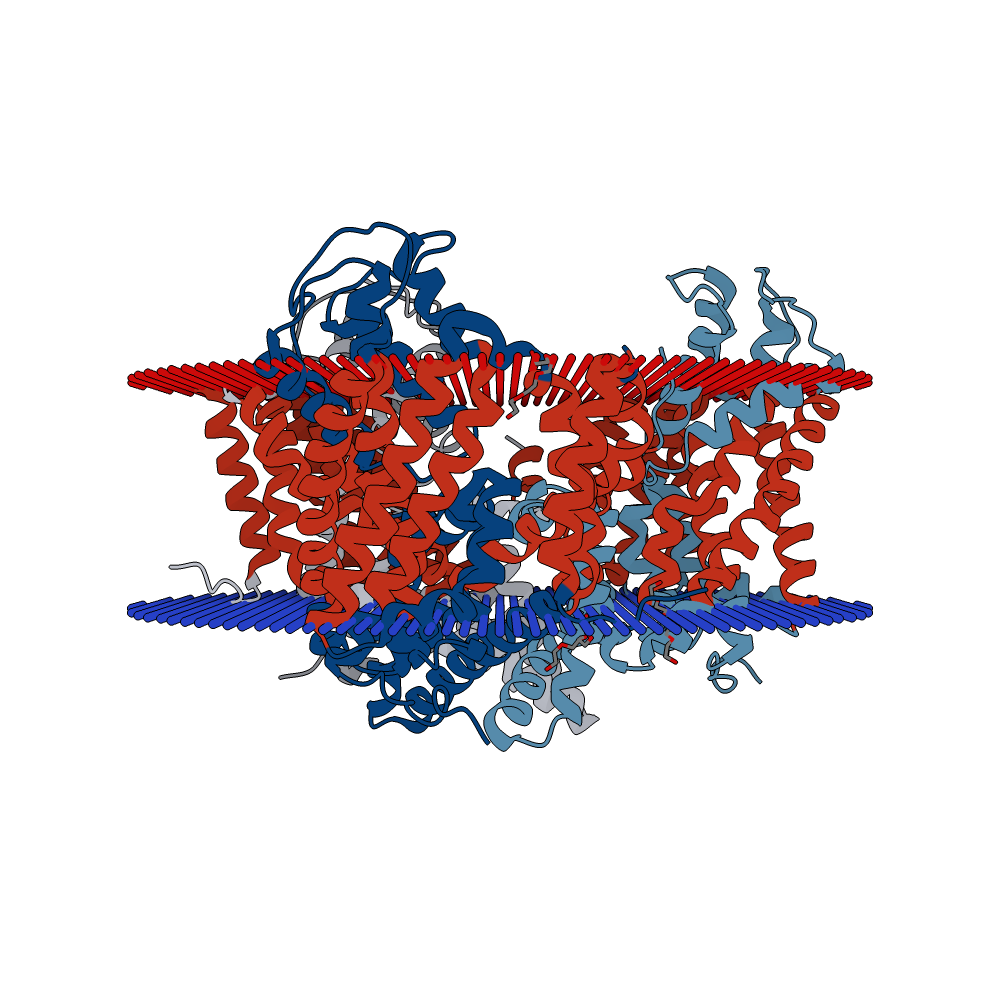
File:T--goettingen--BBa K2586002 structureGltP.png Crystal structure of a substrate-free aspartate transporter.(1,000 × 1,000 (210 KB)) - 10:41, 24 September 2018


