Difference between revisions of "Part:BBa K4712062"
(remove tags) |
|||
| Line 18: | Line 18: | ||
<table><tr><th>Reagent</th><th>Stock Concentration</th><th>Volume Added(μL)</th></tr><tr><td>Forward Primer</td><td>10μM</td><td>1</td></tr><tr><td>Reverse Primer</td><td>10μM</td><td>1</td></tr><tr><td>Rehydration Buffer (2X)</td><td></td><td>10</td></tr><tr><td>DNA Template</td><td>10nM/L</td><td>2</td></tr><tr><td>ddH2O</td><td></td><td>To 18</td></tr><tr><td>Starter (10X)</td><td></td><td>2</td></tr></table> | <table><tr><th>Reagent</th><th>Stock Concentration</th><th>Volume Added(μL)</th></tr><tr><td>Forward Primer</td><td>10μM</td><td>1</td></tr><tr><td>Reverse Primer</td><td>10μM</td><td>1</td></tr><tr><td>Rehydration Buffer (2X)</td><td></td><td>10</td></tr><tr><td>DNA Template</td><td>10nM/L</td><td>2</td></tr><tr><td>ddH2O</td><td></td><td>To 18</td></tr><tr><td>Starter (10X)</td><td></td><td>2</td></tr></table> | ||
| + | |||
| + | https://static.igem.wiki/teams/4712/wiki/result/fig1c.png | ||
| + | |||
| + | Electrophoresis of RPA Primer Screening for Neisseria meningitidis (NME) | ||
| + | |||
| + | https://static.igem.wiki/teams/4712/wiki/result/fig3b.png | ||
| + | |||
| + | The figure above correspond to the fluorescence intensity of crRNA targeting Neisseria meningitidis after CRISPR reaction under Bright and UV illumination. The figure utilize pseudocolor to facilitate analysis using software (Image Lab 6). No crRNA is added into negative control. The concentration of DNA template is 10nM/L. | ||
| + | |||
| + | https://static.igem.wiki/teams/4712/wiki/result/fig3a.png | ||
| + | |||
| + | The linear graphs sequentially correspond to the efficiency verification of crRNAs targeting the DNA sequences of Neisseria meningitidis pathogen. No crRNA is added into negative control. The concentration of DNA template is 10nM/L. | ||
| + | |||
| + | https://static.igem.wiki/teams/4712/wiki/result/fig10c-left.png | ||
| + | https://static.igem.wiki/teams/4712/wiki/result/fig10c-right.png | ||
| + | |||
| + | Linear graphs and figure correspond to the fluorescence intensity of crRNAs targeting NME after one-tube reaction of RPA and CRISPR under Bright and UV illumination. The figures utilize pseudocolor to facilitate analysis using software (Image Lab 6). No crRNA is added into negative control. The concentration of DNA template is 10nM/L. | ||
| + | |||
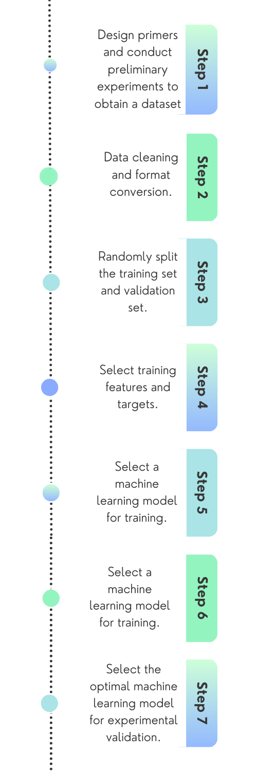
| + | https://static.igem.wiki/teams/4712/wiki/engineering/engineering/fig1.png | ||
Revision as of 12:23, 12 October 2023
NME-F4
The primers were designed using NCBI BLAST and SanpGene to achieve efficient and specific amplification. This primer in RPA serves as the initial binding point for the amplification process, ensuring the specificity of the reaction by targeting the desired Neisseria meningitidis DNA or RNA sequences. The primers provided data for mathematical modeling for further primer design.
Sequence and Features
- 10COMPATIBLE WITH RFC[10]
- 12COMPATIBLE WITH RFC[12]
- 21COMPATIBLE WITH RFC[21]
- 23COMPATIBLE WITH RFC[23]
- 25COMPATIBLE WITH RFC[25]
- 1000COMPATIBLE WITH RFC[1000]
| Reagent | Stock Concentration | Volume Added(μL) |
|---|---|---|
| Forward Primer | 10μM | 1 |
| Reverse Primer | 10μM | 1 |
| Rehydration Buffer (2X) | 10 | |
| DNA Template | 10nM/L | 2 |
| ddH2O | To 18 | |
| Starter (10X) | 2 |

Electrophoresis of RPA Primer Screening for Neisseria meningitidis (NME)

The figure above correspond to the fluorescence intensity of crRNA targeting Neisseria meningitidis after CRISPR reaction under Bright and UV illumination. The figure utilize pseudocolor to facilitate analysis using software (Image Lab 6). No crRNA is added into negative control. The concentration of DNA template is 10nM/L.

The linear graphs sequentially correspond to the efficiency verification of crRNAs targeting the DNA sequences of Neisseria meningitidis pathogen. No crRNA is added into negative control. The concentration of DNA template is 10nM/L.


Linear graphs and figure correspond to the fluorescence intensity of crRNAs targeting NME after one-tube reaction of RPA and CRISPR under Bright and UV illumination. The figures utilize pseudocolor to facilitate analysis using software (Image Lab 6). No crRNA is added into negative control. The concentration of DNA template is 10nM/L.