
Difference between revisions of "Part:BBa K4645001"
Ruixuan Zhu (Talk | contribs) |
Ruixuan Zhu (Talk | contribs) |
||
| Line 3: | Line 3: | ||
<partinfo>BBa_K4645001 short</partinfo> | <partinfo>BBa_K4645001 short</partinfo> | ||
| − | Velocimmune mice were immunized with recombinant dimeric Fel D 1 and produced polyclonal antibodies [1]. Among the resulting polyclonal antibodies, a monoclonal antibody named REGN1908 was found to be the most effective in blocking the immune response to the recombinant dimeric Fel | + | Velocimmune mice were immunized with recombinant dimeric Fel D 1 and produced polyclonal antibodies [1]. Among the resulting polyclonal antibodies, a monoclonal antibody named REGN1908 was found to be the most effective in blocking the immune response to the recombinant dimeric Fel d 1. Through a literature review, we discovered both light chain and heavy chain genes of REGN1908. By fusing the C-terminus of the light chain with the N-terminus of the heavy chain using a rigid linker (GGGGS)<sub>3</sub>, an scFv was obtained. An OmpA signal peptide was fused to the N-terminus of the scFv to help it secrete into the periplasmic space, where it can more effectively neutralize the antigen. |
===Protein Molecular Structures=== | ===Protein Molecular Structures=== | ||
Revision as of 00:24, 12 October 2023
NeuA: scFv ( single-chain fragment variable)that inhibits the immune response to the feline
Velocimmune mice were immunized with recombinant dimeric Fel D 1 and produced polyclonal antibodies [1]. Among the resulting polyclonal antibodies, a monoclonal antibody named REGN1908 was found to be the most effective in blocking the immune response to the recombinant dimeric Fel d 1. Through a literature review, we discovered both light chain and heavy chain genes of REGN1908. By fusing the C-terminus of the light chain with the N-terminus of the heavy chain using a rigid linker (GGGGS)3, an scFv was obtained. An OmpA signal peptide was fused to the N-terminus of the scFv to help it secrete into the periplasmic space, where it can more effectively neutralize the antigen.
Protein Molecular Structures

Design
The plasmid we designed consists of T7 promoter, lac operator, RBS, NeuA coding sequence, His tag, and T7 terminator, which are arranged in an order on a pET28 backbone. We aim to induce the transcription of the downstream NeuA by adding the IPTG to initiate the expression. The protein will then be purified and block activity to FelD was tested by blocking ELISA. We determine the binding effect by coating FelD in the microtiter plates then incubating NeuA and finally measuring the absorbance.

Materials and Method
1.Expression and Purification
1) Plasmid pET-28a(+)-NeuA (with His tag) is transformed to Escherichia coli BL21(DE3). The E. coli strain is cultured in LB medium containing 50 μg/mL kanamycin.
2) When the optical density of the cultured bacteria reached approximately 0.6, IPTG was added to the final concentration 2 mM. And the bacteria were induced at 18℃ overnight. The harvested bacteria are resuspended with a binding buffer (Sangon Biotech, Shanghai, China), and then the bacteria are lysed by ultrasonication. Purification is performed following the instructions of Ni-NTA SefinoseTM Resin (Sangon Biotech, Shanghai, China).
2.Blocking Elisa
1) Microtiter plates were coated overnight at 4 ℃ with FelD.
2) Plates were blocked with 1% BSA for 1 h at RT.
3) Adding 200 μl ELISA Washing Buffer to wash.
4) Different combinations of proteins(NeuA, NeuB, NeuA-NeuB, ClyR) were mixed on the microtiter plates for 90 min at 37℃.
5) Adding 200 μl ELISA Washing Buffer to wash 2 time.
6) Incubating 100 μl Standard IgE sample (25 ng/μl) or 100 μl sera from allergic donors which was diluted 5-fold at 37℃ for 90 min.
7) Incubating 100 μl biotin-labeled IgE antibody at 37℃ for 1 h.
8) Adding 350 μl ELISA Washing Buffer to wash 4 time.
9) Adding 100 μl HRP-conjugated Streptavidin at 37℃ for 30 min.
10) Adding 300 μl ELISA Washing Buffer to wash 4 time.
11) Adding 90 μl color developer (dark) at 37℃ for 15 min.
12) Adding 50 μl termination solution and measuring OD value immediately with microplate reader at 450 nm wavelength.
Result
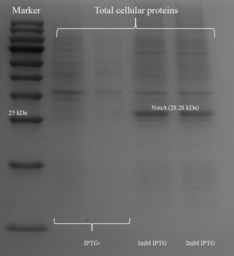
The molecular weight of NeuA is around 28.28 kDa. Thus, the result of SDS-PAGE above indicating that we had the expression of NeuA in our chassis E. coli BL21(DE3) (Figure 3).

Then we successfully expressed and purified the NeuB in the E. coli (Figure 4).
To verify NeuA has the ability to bind to FelD and prevent IgE engagement, We used blocking ELISA to test the blocking activity of different proteins. Compared with the control groups, absorbance at 450 nm of NeuA has an obvious decrease. Additionally, the combination of NeuA and NeuB have better blocking effect (Figure 5). The results of our experiment showed that NeuA has a blocking effect on antigens, and it has a better blocking effect when used together with NeuB. Due to the limited number of sera from allergic donors, we could not have sufficient materials for pre-experiments to reduce absorbance, but the high difference was enough to indicate that our scFvs have blocking activity.

Reference
[1] Orengo JM, Radin AR, Kamat V, Badithe A, Ben LH, Bennett BL, Zhong S, Birchard D, Limnander A, Rafique A, Bautista J, Kostic A, Newell D, Duan X, Franklin MC, Olson W, Huang T, Gandhi NA, Lipsich L, Stahl N, Papadopoulos NJ, Murphy AJ, Yancopoulos GD. Treating cat allergy with monoclonal IgG antibodies that bind allergen and prevent IgE engagement. Nat Commun. 2018 Apr 12;9(1):1421.
Sequence and Features
- 10INCOMPATIBLE WITH RFC[10]Illegal EcoRI site found at 315
Illegal SpeI site found at 75 - 12INCOMPATIBLE WITH RFC[12]Illegal EcoRI site found at 315
Illegal SpeI site found at 75 - 21INCOMPATIBLE WITH RFC[21]Illegal EcoRI site found at 315
Illegal XhoI site found at 693 - 23INCOMPATIBLE WITH RFC[23]Illegal EcoRI site found at 315
Illegal SpeI site found at 75 - 25INCOMPATIBLE WITH RFC[25]Illegal EcoRI site found at 315
Illegal SpeI site found at 75 - 1000COMPATIBLE WITH RFC[1000]