Difference between revisions of "Part:BBa K4140007"
Ahmed Mattar (Talk | contribs) (→Experimental Characterization) |
(→Part Description) |
||
| Line 5: | Line 5: | ||
==Part Description== | ==Part Description== | ||
| − | KP-SP is a secreted signal peptide at the N-terminus of the reporter protein that enables | + | KP-SP is a secreted signal peptide at the N-terminus of the reporter protein that enables its secretion extracellularly with high efficiency which indicates the versatility of this signal peptide and its significant potential in heterologous protein expression. this peptide comes from E.Coli as well as actinomycetes and can be secreted under normal instances |
==Usage== | ==Usage== | ||
Revision as of 18:52, 11 October 2022
kp-sp
Part Description
KP-SP is a secreted signal peptide at the N-terminus of the reporter protein that enables its secretion extracellularly with high efficiency which indicates the versatility of this signal peptide and its significant potential in heterologous protein expression. this peptide comes from E.Coli as well as actinomycetes and can be secreted under normal instances
Usage
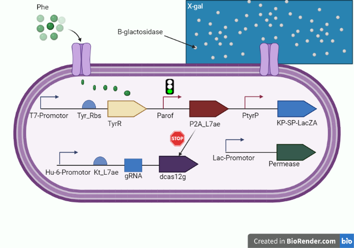
To transmit the response of our circuit to high levels of phenylalanine from intracellular to extracellular we linked Kp Sp signal at the N-terminus of the PAH for transmitting it extracellularly to hydroxylate excess phenylalanine into tyrosine in the therapeutic circuit and to export beta-galactosidase extracellularly to turn the color into blue once bound to its x-gal substrate. as shown in figure 1 and 2.

Figure 1. (shows the tagging of LacZ alpha with kp-sp in the diagnostic whole cell-based biosensor.)

Figure 2. (shows the tagging of PAH with kp-sp in the therapeutic E.coli-based system.)
Characterization by mathematical modeling
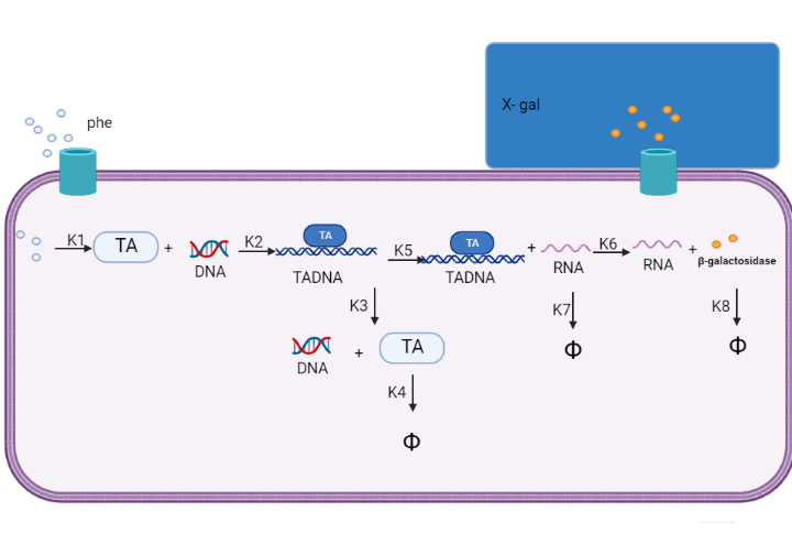
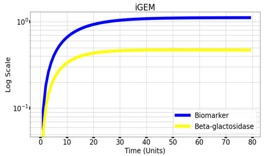
We are using mathematical modeling to detect the increased level of phenylalanine (phe) in phenylketonuria patients in our diagnostic platform. It depends on a whole cell-based biosensor through a cascade of reactions to finally end by formation of β-galactosidase that turns the color into blue once bound to its substrate (X-gal) as mentioned in figure (4) and graph (1).

Figure (4) represents the cascade of reactions in whole cell-based biosensor model.

Graph(1) illustrates a direct relation between biomarker and beta-galactosidase ,so as the biomarker increases, the released amount of beta-galactosidase increases till it reaches constant value after about 30 time units. Therefore, the maximum amount of the biomarker releases the maximum amount of beta-galactosidase.
Experimental Characterization
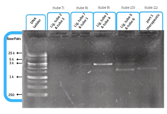
This figure shows an experimental characterization of this part as it's validated through gel electrophoresis as it is in lane 3. The running part (ordered from IDT) included KP-SP and lacZ alpha.
References
Sequence and Features
- 10COMPATIBLE WITH RFC[10]
- 12COMPATIBLE WITH RFC[12]
- 21COMPATIBLE WITH RFC[21]
- 23COMPATIBLE WITH RFC[23]
- 25COMPATIBLE WITH RFC[25]
- 1000COMPATIBLE WITH RFC[1000]

