Difference between revisions of "Part:BBa K3122004"
| Line 1: | Line 1: | ||
__NOTOC__ | __NOTOC__ | ||
| − | <partinfo>BBa_K3122004 | + | <partinfo>BBa_K3122004 short</partinfo> |
This transcriptional unit includes the following parts: | This transcriptional unit includes the following parts: | ||
Latest revision as of 00:51, 22 October 2019
Constitutive medium promoter + strong RBS + AEGIS's lamB display system + 6xHis Tag + Terminator
This transcriptional unit includes the following parts:
- BBa_K3122003: Promoter J23107 adapted to type IIS assembly
- BBa_K2656009: RBS B0030 adapted to type IIS assembly
- BBa_K3122002: coding sequence. This composite part is built with BBa_K3122000 (LamB protein) and BBa_K3122001 (6xHis tag) creating our AEGIS' lamB display system exposing 6xHis Tag.
- BBa_K2656026: Terminator B0015 adapted to type IIS assembly
To see the design of this part go to the Design tab.
Description
This part was used as a cell purification system for the SELEX procedure. Histidines may bind to nickel magnetic beads, allowing the separation and retention of the cells under a magnetic field.
We decided to express a six-unit histidine tag in our LamB display system, in the permissive loop of LamB . The peculiarity of this tag is that histidines bind strongly to nickel beads subjected to a magnetic field. Since histidine is a polar amino acid, negatively charged (due to its multiple amine groups) in a wide range of pH, it can easily interact with ions of metallic particles (Thermofisher, 2019). Using this approach, we suggest that by expressing enough histidine tags in the membrane of our target cells we would be able to induce conjugation between magnetic beads and cells when incubated together in the magnetic module. For further information, visit our Robo-SELEX page .
Besides, by using this promoter of medium strength, we ensure the expression of our protein without overburdening the cell membrane, and therefore, withput killing the cell.
Characterization
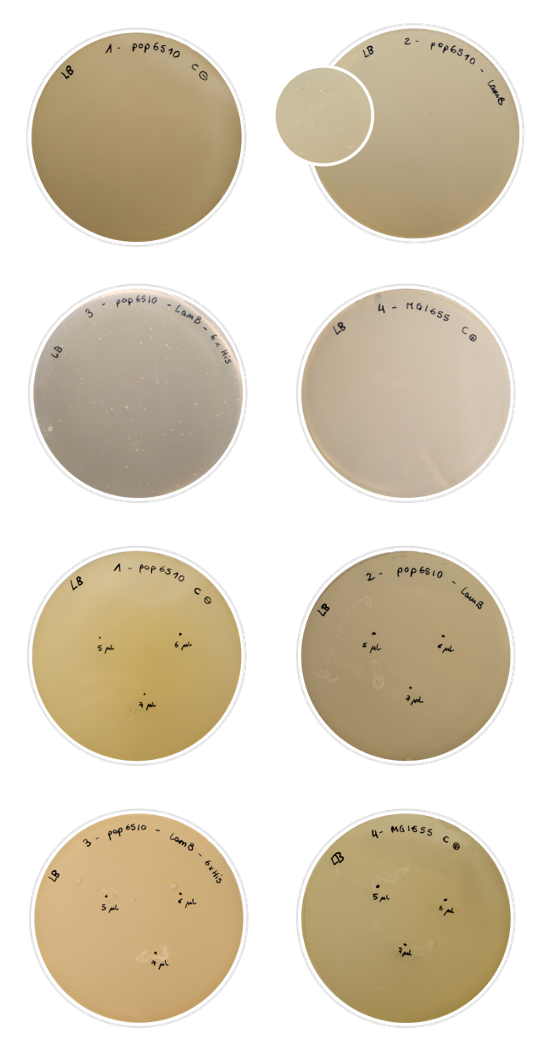
For this characterization, we chose to perform a rather simple test that nonetheless gives conclusive results: the Lambda phage assay. The Lambda phage is a bacteriophage which infects the cell using the LamB protein. We used pop6510 cell line, which does not express LamB. Therefore, if there is no expression of our constructs, the phage cannot infect the cell and there will not be any lysis. On the other hand, if the Lamb display system is expressed properly, the phage will be able to infect the cell, and lysis spots shall appear in the Petri dish.
In this experiment, we characterized the expression in the outer membrane of the LamB protein both with the 6xHis tag and without. We aimed to tell if we had achieved a correct expression of the protein, and if the addition of the His-tag into the permissive loop would compromise this expression.
To prepare the test, we first dabbed the white colonies and made a liquid inoculum. The grown inoculum was treated as explained in the Fague lysis protocol, and then we conducted two different assays:
- Plating in LB agar plates a mix of soft-agar and the result of an incubation of 100μl of the liquid inoculum with 100 μl of phage.
- Plating soft-agar with the bacteria inoculum in LB agar plates with known droplets of Lambda phage: 5,6 and 7 μl in three punctual locations in the Petri dish.
We used regular MG1655 E. coli cells (expressing LamB normally) as positive control, and our pop6510 cells as negative control. The results obtained were positive:
- In the first assay, negative control plate was covered with a bed of cells and positive control plate was clean of cells, as expected. About problem samples, we could observe very few colonies in the petri dish, meaning that the majority of the cells were expressing LamB and therefore lysis has occurred in a high percentage of cells. In the second assay, similar good results were obtained: bald patches appeared in the zone where the Lambda phage was inoculated in the case of problem samples and positive control, while negative control had grown generously.
- In conclusion, the assays could be confirmed as successful. The results explained above were very similar in both constructions, confirming that the introduction of the Tag does not affect the correct expression of LamB in the E. coli outer membrane. However, this result should not be extrapolated to every tag. Bigger or longer tags may lead to misleading results, making this assay unuseful to check its presence.
Characterization of Histidine-based separation
Histidines are used as base of the separation system of our Robo-SELEX protocol. Several alternative strategies were designed, all based on the same concept: the LamB - 6xHis tag functions as an anchoring system, so the cell is retained while aptamers are eluted.
- First strategy: magnetic resin.
- Second strategy: magnetic beads. Following the principle explained above, we tried the same protocol with a different material, this time using cobalt magnetic beads to bind to the Histidine Tag, expressed throughout the surface of E. cholira (J. Kim, 2007).
Results
- We conducted a first assay with the magnetic resin using the maximum values for all the variables we wanted to optimize (initial concentration of cells and incubation time), and as the proportion of cells found after plating the dilutions we were unable to drag or retain enough cells. After analyzing our results, we concluded that due to the small size of the particles in the resin, they would be too compact with a pore size too narrow to allow let the cell access to the inside of the resin matrix.
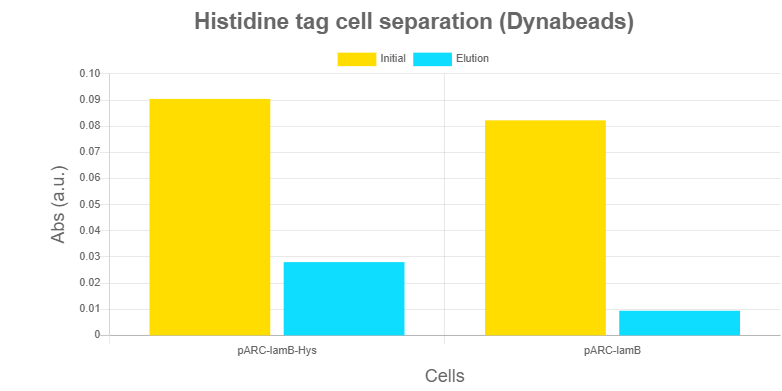
- We attempted to separate the cells with the cobalt magnetic beads. We thought that the individuality of the beads, which are present in the solution without aggregating in a small-pore-sized resin, would correct those problems. An assay was conducted as a proof of concept, comparing the absorbance between Eppendorfs that suffer E. cholira separation with and without expressing the Histidine tag. So, after performing the assay in the OT2, we measured absorbance at 640nm of the resulting 96 well-plate.
The results obtained in this assay showed a significant difference between the control cells pop6510 harbouring vector pARK1-LamB (without expressing the anchor systems) and the pop6510 harbouring pARK1LamB-6xHis. With this, we confirmed that there was an interaction between the Tag and the cobalt beads. After analyzing the results obtained in the assay we saw that the efficiency of the process was around 10%, which was not enough to conduct a proper SELEX protocol due to the number of aptamers in each SELEX round that we would lose in the remaining 90% of the cells.
How to use it
This is an example of how this new display system that we created can be used by future iGEM teams on their MoClo reactions with the desired tag to create level 1 constructs. The only requirement is to adapt the tag sequence to the new scars designed, and fulfill the standard for the rest of parts.
- 10COMPATIBLE WITH RFC[10]
- 12INCOMPATIBLE WITH RFC[12]Illegal NheI site found at 11
Illegal NheI site found at 34 - 21COMPATIBLE WITH RFC[21]
- 23COMPATIBLE WITH RFC[23]
- 25INCOMPATIBLE WITH RFC[25]Illegal AgeI site found at 212
Illegal AgeI site found at 1301 - 1000COMPATIBLE WITH RFC[1000]
Judging
This transcriptional unit has been used to characterize the existing BBa_J23107 Promoter basic part (adapted to type IIS gramar in the new part BBa_K3122003), BBa_K2656009 RBS basic part and BBa_K2656026 Terminator basic part.
References
J. Kim, C. Valencia, R. Liu and W. Lin, "Highly-Efficient Purification of Native Polyhistidine-Tagged Proteins by Multivalent NTA-Modified Magnetic Nanoparticles", Bioconjugate Chemistry, vol. 18, no. 2, pp. 333-341, 2007. Available:10.1021/bc060195l.


