Difference between revisions of "Part:BBa K3114000"
(→Characterization) |
|||
| Line 38: | Line 38: | ||
[[File:T--Calgary--SPGraphRegistry.png|650px|thumb|center|Figure 3. Relative concentration of 6GIX protein obtained by performing a periplasmic isolation after secretion using various fused signal peptides. 50 ml cultures were grown to OD<sub>600</sub>=0.4 then induced with 100 mM IPTG for 8 hours at 37°C before the periplasmic isolation was performed. Genetic constructs for 6GIX without a fused signal peptide and the empty vector were used as controls. Values were obtained by spectrophotometric determination of protein concentration, and represent the mean of two replicates.]] | [[File:T--Calgary--SPGraphRegistry.png|650px|thumb|center|Figure 3. Relative concentration of 6GIX protein obtained by performing a periplasmic isolation after secretion using various fused signal peptides. 50 ml cultures were grown to OD<sub>600</sub>=0.4 then induced with 100 mM IPTG for 8 hours at 37°C before the periplasmic isolation was performed. Genetic constructs for 6GIX without a fused signal peptide and the empty vector were used as controls. Values were obtained by spectrophotometric determination of protein concentration, and represent the mean of two replicates.]] | ||
| + | |||
| + | The SDS-PAGE gel below shows the protein recovered from the periplasm for each of our genetic constructs as per our periplasmic protein isolation [https://2019.igem.org/Team:Calgary/Experiments protocol.] The construct with the MalE signal peptide showed the strongest band of the correct size, but bands are also visible for the DsbA and PhoA signal peptide samples. | ||
| + | |||
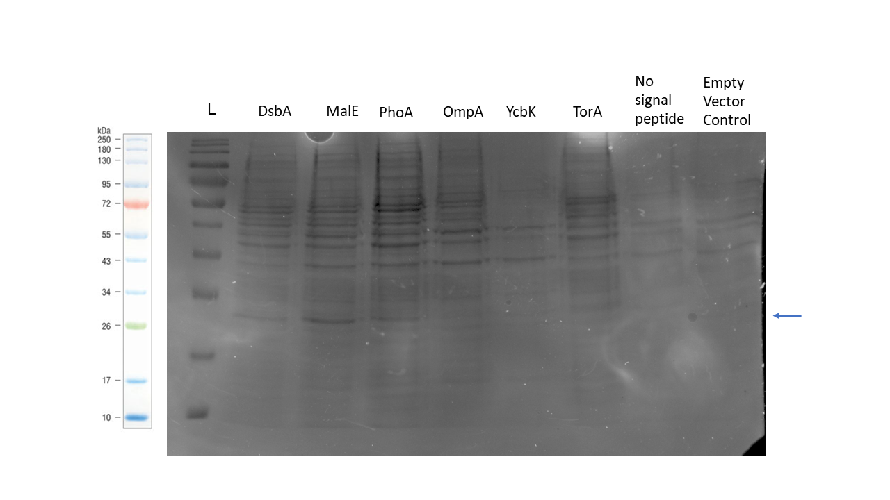
| + | [[Image:T--Calgary--SPgel.png|700px|thumb|center|Figure 4. SDS-PAGE gel showing periplasmic purification of 6GIX with six different signal peptides, no signal peptide, and an empty vector control. The marker used is the NEB colour protein standard. The arrow denotes correct band size of 21 kDa for the 6GIX protein.]] | ||
===Sequence and Features=== | ===Sequence and Features=== | ||
Revision as of 05:27, 21 October 2019
Signal peptide from DsbA (secretion tag)
Usage and Biology
The DsbA signal peptide is a 19-amino acid sequence which targets fused proteins to the Sec secretion pathway in E. coli. The fused protein is not folded until it is secreted to the periplasm. The signal peptide is cleaved after residue 19 by signal peptidase after secretion (recognition sequence: Ala-X-Ala).
Translocation of recombinant proteins to the periplasm can be an effective strategy for increasing protein yield by mitigating inclusion body formation and for simplifying purification procedures in E. coli (Sørensen and Mortensen, 2005). Many different signal peptide sequences have been characterized in literature. However, there are multiple factors that influence the ability of a particular signal peptide to target the recombinant protein of interest to the periplasm (Freudl, 2018). The optimal signal peptide candidate must therefore be determined experimentally. As such, iGEM Calgary sought to create a set of signal peptide parts so that teams may test multiple candidates in their genetic constructs to obtain optimal secretion. The parts were designed to be Golden Gate-compatible based on the MoClo standard for ease in creating the genetic constructs required to screen for the optimal candidate (Weber et al., 2011). They are also compatible with the BioBrick RFC[10] standard.
This signal peptide sequence originates from the DsbA protein in E. coli, which is a periplasmic disulfide bond oxidoreductase (Schierle et al., 2003). Other signal peptides within this collection include:
- MalE signal peptide (BBa_K3114001)
- OmpA signal peptide (BBa_K3114002)
- PhoA signal peptide (BBa_K3114003)
- TorA signal peptide (BBa_K3114005)
- YcbK signal peptide (BBa_K3114004)
Design
When designing this part and the rest of our collection, we were interested in creating parts that could be used in Golden Gate assembly right out of the distribution kit without the need to first domesticate them in a Golden Gate entry vector. As such, these parts are not compatible with the iGEM Type IIS RFC[1000] assembly standard because we included the BsaI restriction site and MoClo standard fusion site in the part’s sequence.
As per the MoClo standard, the 5’ signal peptide fusion sequence included in this part is AATG, and the 3’ signal peptide fusion sequence is AGGT.
This part includes a start codon. A Gly-Gly spacer sequence was added to the end of the signal peptide sequence in order to ensure that the fused protein of interest will be in-frame following Golden Gate assembly.
This sequence has been codon optimized for high expression in E. coli.
Characterization
Each of the signal peptides in this collection was fused to the water-soluble chlorophyll binding protein 6GIX in a genetic circuit under the control of a T7 inducible promoter. For more information, please see iGEM Calgary's parts page.
The graph below represents the concentration of 6GIX isolated from the periplasm of for each culture. The results suggest that the DsbA, MalE, PhoA, and TorA signal peptides are our most effective candidates for localization of 6GIX to the periplasmic space. Concentrations were determined using spectrophotometry.

The SDS-PAGE gel below shows the protein recovered from the periplasm for each of our genetic constructs as per our periplasmic protein isolation protocol. The construct with the MalE signal peptide showed the strongest band of the correct size, but bands are also visible for the DsbA and PhoA signal peptide samples.
Sequence and Features
- 10COMPATIBLE WITH RFC[10]
- 12COMPATIBLE WITH RFC[12]
- 21COMPATIBLE WITH RFC[21]
- 23COMPATIBLE WITH RFC[23]
- 25COMPATIBLE WITH RFC[25]
- 1000INCOMPATIBLE WITH RFC[1000]Illegal BsaI site found at 1
Illegal BsaI.rc site found at 73
References
Freudl, R. (2018, March 29). Signal peptides for recombinant protein secretion in bacterial expression systems. Microbial Cell Factories. BioMed Central Ltd. https://doi.org/10.1186/s12934-018-0901-3
Schierle, C. F., Berkmen, M., Huber, D., Kumamoto, C., Boyd, D., & Beckwith, J. (2003). The DsbA signal sequence directs efficient, cotranslational export of passenger proteins to the Escherichia coli periplasm via the signal recognition particle pathway. Journal of Bacteriology, 185(19), 5706–5713. https://doi.org/10.1128/JB.185.19.5706-5713.2003
Sørensen, H. P., & Mortensen, K. K. (2005). Advanced genetic strategies for recombinant protein expression in Escherichia coli. Journal of Biotechnology, 115(2), 113–128. https://doi.org/10.1016/j.jbiotec.2004.08.004
Weber, E., Engler, C., Gruetzner, R., Werner, S., & Marillonnet, S. (2011). A modular cloning system for standardized assembly of multigene constructs. PLoS ONE, 6(2). https://doi.org/10.1371/journal.pone.0016765


