Difference between revisions of "Part:BBa K1991009"
| Line 9: | Line 9: | ||
[[File:2016Mingdao Proof2.jpg|300px|thumb|left]] | [[File:2016Mingdao Proof2.jpg|300px|thumb|left]] | ||
| + | |||
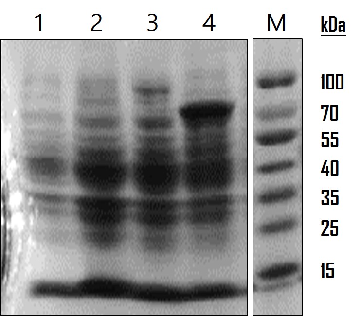
| + | <p>Figure 1: AOX protein analysis. SDS-PAG and Coomassie Blue staining were used to observe protein expression level. Lane 1: wild-type E. coli as a mock control; Lane 2: LO outer membrane protein ([https://parts.igem.org/Part:BBa_K1991007 BBa_K1991007]) (17 kDa) expression in E. coli; Lane 3: LO-AOX fusion protein [https://parts.igem.org/Part:BBa_K1991009 BBa_K1991009]) (91 kDa) expression in E. coli.; Lane 4: AOX protein [https://parts.igem.org/Part:BBa_K1991003 BBa_K1991003]) (79 kDa) expression in E. coli.</p> | ||
Revision as of 19:37, 19 October 2016
Pcons-RBS-LO-AOX2-His
DISPLAYING AOX ENZYME ON THE CELL SURFACE OF E. COLI
A protein can be displayed on the cell surface of E. coli by fusing to Lpp-OmpA. Lipoprotein ([http://www.uniprot.org/uniprot/P69776 Lpp]) is a major outer membrane of E. coli which interacts with the peptidoglycan to maintain the structure and function of cell membrane. Another transmembrane protein called outer membrane protein A ([http://www.uniprot.org/uniprot/P0A910 OmpA]) is involved in bacterial conjugation and phage infection. A study showed that Lpp-OmpA hybrid can direct the heterologous protein GFP to the external surface of E. coli (Enzyme Microb Technol. 2001). Bacillus lipase (J Microbiol. 2014) and Fungi xylanase (Curr Microbiol. 2015) were demonstrated to be displayed on the cell surface of E. coli and maintained the functional enzyme activities. In iGEM 2016 MINGDAO's project, AOX (alcohol oxidase) was displayed on bacterial surface by fusing with Lpp-OmpA, which was proved by protein analysis, enzyme activity assay and TMB assay
PROTEIN EXPRESSION ANALYSIS
To analyze the AOX gene expression, we run on a SDS-PAGE gel and observed by Coomassie Blue staining. The overnight-cultured E. coli were centrifuged and lysed with Lysis Buffer (12.5 mM Tris pH 6.8, 4% SDS). The resulting lysates were subjected to SDS-PAGE with a 10% polyacrylamide gel. The gel was stained with 0.25% Coomassie Brilliant Blue R250 for 2 hours and destained until the protein bands were clear. As the data showed in Figure 1, LO protein was expressed at around the estimated molecular weight of 17 kDa, LO-AOX fusion protein at 91 kDa and AOX protein at 79 kDa. The protein expression level was low compared to AOX expression but significant compared to WT and LO. However, so far we cannot confirm whether LO fusion protein is able to direct AOX or GFP proteins displayed on the cell surface of E. coli. We’re planning to do a subcellular fractionation to separate the outer membrane proteins for analysis in the future.
Figure 1: AOX protein analysis. SDS-PAG and Coomassie Blue staining were used to observe protein expression level. Lane 1: wild-type E. coli as a mock control; Lane 2: LO outer membrane protein (BBa_K1991007) (17 kDa) expression in E. coli; Lane 3: LO-AOX fusion protein BBa_K1991009) (91 kDa) expression in E. coli.; Lane 4: AOX protein BBa_K1991003) (79 kDa) expression in E. coli.
ENZYME ACTIVITY ASSAY
TMB ASSAY PERFORMED (BY NCTU-FORMOSA 2016)
Sequence and Features
- 10COMPATIBLE WITH RFC[10]
- 12INCOMPATIBLE WITH RFC[12]Illegal NheI site found at 7
Illegal NheI site found at 30
Illegal NotI site found at 2498 - 21INCOMPATIBLE WITH RFC[21]Illegal BamHI site found at 497
Illegal XhoI site found at 2507 - 23COMPATIBLE WITH RFC[23]
- 25INCOMPATIBLE WITH RFC[25]Illegal NgoMIV site found at 452
- 1000COMPATIBLE WITH RFC[1000]

