Difference between revisions of "Part:BBa K1420005"
| Line 9: | Line 9: | ||
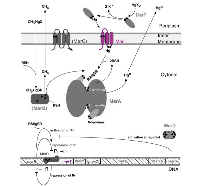
'''Figure 1.''' Model of transmembrane mercuric binding protein MerT. The symbol • indicates a cysteine residue. RSH indicates cytosolic thiol redox buffers such as glutathione. Figure 1 shows the interactions of MerT, in purple, with mercury compounds and other gene products of ''mer'' operon. (This figure is adapted from "Bacterial mercury resistance from atoms to ecosystems". Reference: ''T. Barkay et al''. FEMS Microbiology Reviews 27 (2003) 355-384.) | '''Figure 1.''' Model of transmembrane mercuric binding protein MerT. The symbol • indicates a cysteine residue. RSH indicates cytosolic thiol redox buffers such as glutathione. Figure 1 shows the interactions of MerT, in purple, with mercury compounds and other gene products of ''mer'' operon. (This figure is adapted from "Bacterial mercury resistance from atoms to ecosystems". Reference: ''T. Barkay et al''. FEMS Microbiology Reviews 27 (2003) 355-384.) | ||
<p></p> | <p></p> | ||
| − | |||
| − | |||
<b> Protein </b> | <b> Protein </b> | ||
Revision as of 03:02, 17 October 2014
merT, mercuric transport protein
Transmembrane mercuric binding gene merT (0.3Kb) encodes MerT, which transports Hg(II) species from the periplasm through the membrane. MerT and gene merT are highlighted in purple in Figure 1. The lower part of the Figure 1 shows the arrangement of mer genes in the operon, and merT is located upstream of merP, another gene that encodes a mercury transport protein. The gene merT is found in the Serratia marcescens plasmid pDU1358 downstream of the bidirectional promoter of merR, and it is 351 base pairs long.
Figure 1. Model of transmembrane mercuric binding protein MerT. The symbol • indicates a cysteine residue. RSH indicates cytosolic thiol redox buffers such as glutathione. Figure 1 shows the interactions of MerT, in purple, with mercury compounds and other gene products of mer operon. (This figure is adapted from "Bacterial mercury resistance from atoms to ecosystems". Reference: T. Barkay et al. FEMS Microbiology Reviews 27 (2003) 355-384.)
Protein
MerT is a 116-residue (12.4 kDa) transmembrane protein. The exact structure is not known, but the protein is predicted to have three transmembrane helices with a cysteine pair accessible from the periplasmic side and another pair on the cytoplasmic side.
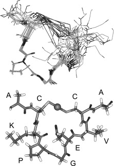
Fig 2. Structure of MerT bound to Hg(II). Top Panel: Images of the 20 lowest energy states that have been superimposed upon each other. Bottom Panel: Lowest energy structure of MerT in which the sphere represents the mercury. Reference: E. Rossy et al. FEBS Lett. (2004) 575, 86–90.
Mechanism
The transport mechanism of ionic mercury from the bacterial cell periplasm to cytosplasm is not completely understood. Hg(II) is transferred from the periplasmic cysteine pair on the first transmembrane helix to the cytoplasmic loop cysteine, where it is finally transferred to a cysteine pair at the N-terminus of the protein. The Hg(II) may go directly to MerA from the N-terminus of MerT or transfer to cytoplasmic low-molecular mass thiols.
Sequence and Features
- 10COMPATIBLE WITH RFC[10]
- 12INCOMPATIBLE WITH RFC[12]Illegal NheI site found at 45
- 21COMPATIBLE WITH RFC[21]
- 23COMPATIBLE WITH RFC[23]
- 25COMPATIBLE WITH RFC[25]
- 1000INCOMPATIBLE WITH RFC[1000]Illegal SapI site found at 30


