Difference between revisions of "Part:BBa K1149050"
| Line 40: | Line 40: | ||
https://static.igem.org/mediawiki/2013/thumb/9/95/Glucose_inhibition.png/450px-Glucose_inhibition.png | https://static.igem.org/mediawiki/2013/thumb/9/95/Glucose_inhibition.png/450px-Glucose_inhibition.png | ||
| − | Glucose inhibition on growth in minimal media on bdh2. The outcome of this growth assay shows that cells grow more in M9WCM but that increasing glucose concentration influences growth in M9WCM while it is unaffected in M9 minimal media. Characterisation was done in M9 and M9WCM. Data points show final time point after 6h growth for each concentration. The arabinose concentration was kept at 6 μM for all samples. Growth was at 37°C with shaking over 6h. Error bars are SEM, n=4. Figure made by Imperial College London 2013 iGEM. | + | '''Glucose inhibition on growth in minimal media on bdh2.''' The outcome of this growth assay shows that cells grow more in M9WCM but that increasing glucose concentration influences growth in M9WCM while it is unaffected in M9 minimal media. Characterisation was done in M9 and M9WCM. Data points show final time point after 6h growth for each concentration. The arabinose concentration was kept at 6 μM for all samples. Growth was at 37°C with shaking over 6h. Error bars are SEM, n=4. Figure made by Imperial College London 2013 iGEM. |
https://static.igem.org/mediawiki/2013/thumb/0/0d/F_glucose_inhibition.png/450px-F_glucose_inhibition.png | https://static.igem.org/mediawiki/2013/thumb/0/0d/F_glucose_inhibition.png/450px-F_glucose_inhibition.png | ||
| − | Glucose inhibition on fluorescence in minimal media on bdh2. The fluorescence of GFP in bdh2 under M9WCM shows significant difference in fluorescence output (p < 0.0001.). This compared to M9 media where fluorescence is almost half. The fluorescence in glucose is quite stable despite the decreased growth witnessed in the growth assay. Characterisation was done in M9 and M9WCM. Data points show final time point after 6h growth for each concentration. The arabinose concentration was kept at 6 μM for all samples. Growth was at 37°C with shaking over 6h. Error bars are SEM, n=4. Figure made by Imperial College London 2013 iGEM. | + | '''Glucose inhibition on fluorescence in minimal media on bdh2.''' The fluorescence of GFP in bdh2 under M9WCM shows significant difference in fluorescence output (p < 0.0001.). This compared to M9 media where fluorescence is almost half. The fluorescence in glucose is quite stable despite the decreased growth witnessed in the growth assay. Characterisation was done in M9 and M9WCM. Data points show final time point after 6h growth for each concentration. The arabinose concentration was kept at 6 μM for all samples. Growth was at 37°C with shaking over 6h. Error bars are SEM, n=4. Figure made by Imperial College London 2013 iGEM. |
| − | Conclusion: The null hypothesis, glucose concentration does not influence growth of MG1655 in M9 minimal (M9M) media and M9 minimal waste conditioned media (M9MWCM) was tested using a two-tailed t-test. This must be rejected because p < 0.0001, thus '''glucose concentration has an influence on growth in M9M and M9MWCM.''' In addition to this, the null hypothesis: glucose concentration does not influence fluorescence in M9 minimal media must be rejected as p < 0.0001.''' Thus glucose concentration influences fluorescence. | + | <span style="color:#f00">'''Conclusion:'''</span> The null hypothesis, glucose concentration does not influence growth of MG1655 in M9 minimal (M9M) media and M9 minimal waste conditioned media (M9MWCM) was tested using a two-tailed t-test. This must be rejected because p < 0.0001, thus '''glucose concentration has an influence on growth in M9M and M9MWCM.''' In addition to this, the null hypothesis: glucose concentration does not influence fluorescence in M9 minimal media must be rejected as p < 0.0001.''' Thus glucose concentration influences fluorescence. |
''' | ''' | ||
Revision as of 23:57, 2 October 2013
Bdh2 (intracellular)
This part expresses Bdh2 3HB dehydrogenase enzyme intracellularly. There is a GFP in operon with the enzyme which can be used to monitor expression levels. The promoter is arabinose inducible pBAD.
http://www.igem.org/wiki/images/f/f0/Reaction_bdh.jpg
Usage and Biology
Characterisation
Growth in LB and fluorescence
Our bdh2 (intracellular) construct contains sfGFP within an operon and therefore fluorescence can be utilised to determine if expression is being induced by addition of Arabinose.

Bdh2 with no pelB secretion tag growth assay. MG1655 with intracellular bdh2 were grown over 6h to gauge the effect on growth when induced. Graph shows that while they reach the same end point, growth is initially faster without induction for BBa_K1149050, though this was not significant as a two-tailed t-test gave p = 0.4930 > 0.05, allowing us to accept the null hypothesis. Growth was at 37°C in LB with shaking. Error bars are SEM, n=4. Figure made by Imperial College London 2013 iGEM.

Bdh2 with no pelB secretion tag induction assay. In addition to testing BBa_K1149050 growth, we looked at induction whereby we see that more sfGFP is produced when induced. Although the promoter is leaky as the curve of intracellular bdh2, as seen with bdh2-pelB is followed tightly by the non-induced cells. Growth was at 37°C in LB with shaking. Error bars are SEM, n=4. Figure made by Imperial College London 2013 iGEM.
Conclusion: There is no growth inhibition caused by induction, nor is there any significant fluorescence induction.
Growth in Minimal media
Arabinose induction in M9 minimal media

Influence of Arabinose Concentration on growth in minimal media on bdh2. This showed that while growth was unaffected in bdh2 with Arabinose induction from 2-10 μM, M9WCM had decreased growth with an optimum at 10 μM. Characterisation was done in both M9 minimal media and M9 minimal [http://2013.igem.org/Team:Imperial_College/Protocols#Waste_Conditioned_Media_.28WCM.29 waste conditioned media]. Data points show final time point after 6h growth for each concentration. 0.4% glucose was used as a carbon source. Growth was at 37°C with shaking over 6h. Error bars are SEM, n=4. Figure made by Imperial College London 2013 iGEM.

Influence of Arabinose Concentration on fluorescence in minimal media on bdh2. The trend shows that fluorescence of GFP in bdh2 is increased in M9 at lower induction concentration than M9WCM, however, the graph requires normalisation to better reflect reality. Characterisation was done in M9 and M9WCM. Data points show final time point after 6h growth for each concentration. 0.4% glucose was used as a carbon source. Growth was at 37°C with shaking over 6h. Error bars are SEM, n=4. Figure made by Imperial College London 2013 iGEM.
Conclusion: The null hypothesis, arabinose concentration does not influence growth of MG1655 in M9 minimal (M9M) media and M9 minimal waste conditioned media (M9MWCM) was tested using a two-tailed t-test. This must be rejected because p < 0.0217, thus arabinose concentration has an influence on growth in M9M and M9MWCM.
Glucose inhibition in M9 minimal media

Glucose inhibition on growth in minimal media on bdh2. The outcome of this growth assay shows that cells grow more in M9WCM but that increasing glucose concentration influences growth in M9WCM while it is unaffected in M9 minimal media. Characterisation was done in M9 and M9WCM. Data points show final time point after 6h growth for each concentration. The arabinose concentration was kept at 6 μM for all samples. Growth was at 37°C with shaking over 6h. Error bars are SEM, n=4. Figure made by Imperial College London 2013 iGEM.

Glucose inhibition on fluorescence in minimal media on bdh2. The fluorescence of GFP in bdh2 under M9WCM shows significant difference in fluorescence output (p < 0.0001.). This compared to M9 media where fluorescence is almost half. The fluorescence in glucose is quite stable despite the decreased growth witnessed in the growth assay. Characterisation was done in M9 and M9WCM. Data points show final time point after 6h growth for each concentration. The arabinose concentration was kept at 6 μM for all samples. Growth was at 37°C with shaking over 6h. Error bars are SEM, n=4. Figure made by Imperial College London 2013 iGEM.
Conclusion: The null hypothesis, glucose concentration does not influence growth of MG1655 in M9 minimal (M9M) media and M9 minimal waste conditioned media (M9MWCM) was tested using a two-tailed t-test. This must be rejected because p < 0.0001, thus glucose concentration has an influence on growth in M9M and M9MWCM. In addition to this, the null hypothesis: glucose concentration does not influence fluorescence in M9 minimal media must be rejected as p < 0.0001. Thus glucose concentration influences fluorescence.
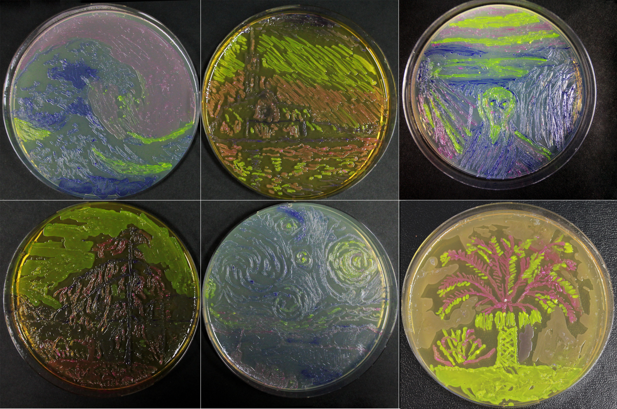
Living Art
In addition to this, we have shown that bacterial paintings are achievable with this amazing biobrick (yellow):

References:
Sequence and Features
- 10COMPATIBLE WITH RFC[10]
- 12INCOMPATIBLE WITH RFC[12]Illegal NheI site found at 125
- 21INCOMPATIBLE WITH RFC[21]Illegal BamHI site found at 65
- 23COMPATIBLE WITH RFC[23]
- 25INCOMPATIBLE WITH RFC[25]Illegal AgeI site found at 181
Illegal AgeI site found at 1126 - 1000COMPATIBLE WITH RFC[1000]
