Difference between revisions of "Part:BBa K4325026"
| Line 21: | Line 21: | ||
<h4>1.Batch screening of pDawn(cI-LVA)-RBS070-LKD-T1 in response to blue light lysis in <i>E. coli</i> TOP10.</h4> | <h4>1.Batch screening of pDawn(cI-LVA)-RBS070-LKD-T1 in response to blue light lysis in <i>E. coli</i> TOP10.</h4> | ||
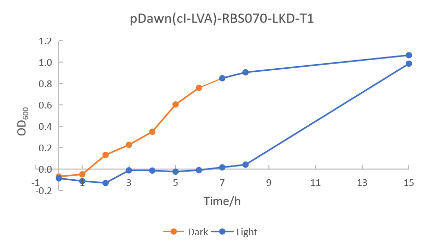
| − | <p>As shown in Figure 2, except for the 11<sup>th</sup> and 13<sup>th</sup> bacterial colonies, other bacterial colonies did not grow under blue light. To further explore the lysis effect of pDawn(cI-LVA)-RBS070-LKD-T1-TOP10, we tracked the OD<sub> | + | <p>As shown in Figure 2, except for the 11<sup>th</sup> and 13<sup>th</sup> bacterial colonies, other bacterial colonies did not grow under blue light. To further explore the lysis effect of pDawn(cI-LVA)-RBS070-LKD-T1-TOP10, we tracked the OD<sub>600</sub> values of pDawn(cI-LVA)-RBS070-LKD-T1-TOP10 and plotted the growth curves diagram. As shown in Figure 3, the lysis effect of pDawn(cI-LVA)-RBS070-LKD-T1-TOP10 meets our expectation in the first eight hours, but it was unstable after fifteen hours.</p> |
[[File:K26 2.png|600px|thumb|center|Figure 2: Growth conditions of <i>E.coli</i>TOP10-pDawn(cI-LVA)-RBS070-LKD-T1 in the dark and under the light.]] | [[File:K26 2.png|600px|thumb|center|Figure 2: Growth conditions of <i>E.coli</i>TOP10-pDawn(cI-LVA)-RBS070-LKD-T1 in the dark and under the light.]] | ||
Revision as of 14:24, 12 October 2022
pDawn-RBS070-LKD-T1
Description
This composite part is a generator containing pDawn(cI-LVA) (BBa_K1075044), RBS070 (BBa_K4325002), LKD (BBa_K4325004) and T1 terminator(BBa_K3033016).
Usage
We inserted the blue light responsive system pDawn(cI-LVA) (BBa_K1075044) and lysis gene LKD (BBa_K4325004) into the pSEVA331 expression vector, which was incorporated into E. coli TOP10 and screened for the bacterial colonies which grew in the dark but did not grow under blue light. Finally, the plasmids containing pDawn(cI-LVA)-RBS070-LKD-T1 were selected and introduced into G. hansenii ATCC53582 by electroporation to verify the responsiveness of pDawn(cI-LVA) to blue light.
Sequence and Features
- 10COMPATIBLE WITH RFC[10]
- 12COMPATIBLE WITH RFC[12]
- 21INCOMPATIBLE WITH RFC[21]Illegal BglII site found at 2171
- 23COMPATIBLE WITH RFC[23]
- 25INCOMPATIBLE WITH RFC[25]Illegal NgoMIV site found at 63
Illegal NgoMIV site found at 195
Illegal NgoMIV site found at 289
Illegal NgoMIV site found at 582
Illegal NgoMIV site found at 1076
Illegal NgoMIV site found at 1094
Illegal NgoMIV site found at 1184
Illegal AgeI site found at 414
Illegal AgeI site found at 1542 - 1000INCOMPATIBLE WITH RFC[1000]Illegal BsaI site found at 1643
Illegal BsaI.rc site found at 525
2022 SZPT-China
Characterization
1.Batch screening of pDawn(cI-LVA)-RBS070-LKD-T1 in response to blue light lysis in E. coli TOP10.
As shown in Figure 2, except for the 11th and 13th bacterial colonies, other bacterial colonies did not grow under blue light. To further explore the lysis effect of pDawn(cI-LVA)-RBS070-LKD-T1-TOP10, we tracked the OD600 values of pDawn(cI-LVA)-RBS070-LKD-T1-TOP10 and plotted the growth curves diagram. As shown in Figure 3, the lysis effect of pDawn(cI-LVA)-RBS070-LKD-T1-TOP10 meets our expectation in the first eight hours, but it was unstable after fifteen hours.
References
[1] Robert Ohlendorf, Roee R. Vidavski, Avigdor Eldar et.al.From Dusk till Dawn: One-Plasmid Systems for Light-Regulated Gene Expression.Journal of Molecular Biology, 08 Jan 2012, 416(4):534-542.
[2] [2]Ceyssens, P.-J. et al. Genomic analysis of Pseudomonas aeruginosa phages LKD16 and LKA1: establishment of the phiKMV subgroup within the T7 supergroup. J. Bacteriol. 188, 6924-31 (2006).


