Difference between revisions of "Part:BBa K3037002"
(→Characterization) |
(→Experiment in Detail) |
||
| Line 57: | Line 57: | ||
===== 1. DNA-binding ability of dCas9 (BBa K3037002)===== | ===== 1. DNA-binding ability of dCas9 (BBa K3037002)===== | ||
| − | <b> | + | <b>a) Materials:</b> |
*100 ng of PCR amplified <span style="font-style: italic;">sry</span> gene | *100 ng of PCR amplified <span style="font-style: italic;">sry</span> gene | ||
| Line 84: | Line 84: | ||
[[File:T--TU_Dresden--EMSA_Primers_BBa_K3037005.png|center|400px|thumb|left|Table 1: Overview of different guide RNAs with the context of the sequence and the PAM sequence]] | [[File:T--TU_Dresden--EMSA_Primers_BBa_K3037005.png|center|400px|thumb|left|Table 1: Overview of different guide RNAs with the context of the sequence and the PAM sequence]] | ||
| − | <b> | + | <b>b) Methods:</b> |
1. We wanted to check if the overall efficiency of mobility shift increases when combinations of guide RNAs are used. | 1. We wanted to check if the overall efficiency of mobility shift increases when combinations of guide RNAs are used. | ||
| Line 95: | Line 95: | ||
| − | <b> | + | <b>3c) (i) Results and Discussion of the 2 hours gel:</b> |
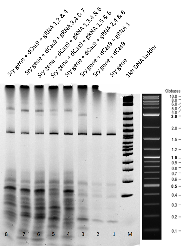
[[File:T--TU_Dresden--EMSA1_BBa_K3037005.png|center|400px|thumb|left|Figure 1: Electrophoretic Mobility Shift Assay (EMSA) after a two hour run using dCas9 with different guideRNAs]] | [[File:T--TU_Dresden--EMSA1_BBa_K3037005.png|center|400px|thumb|left|Figure 1: Electrophoretic Mobility Shift Assay (EMSA) after a two hour run using dCas9 with different guideRNAs]] | ||
| Line 108: | Line 108: | ||
| − | <b> | + | <b>c) (ii)Results and Discussion of the 3 hours gel:</b> |
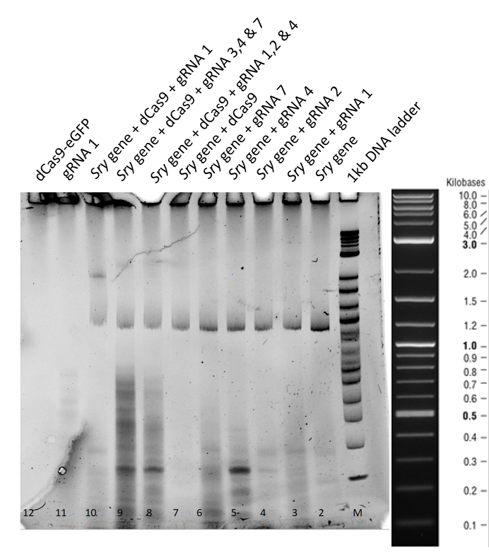
[[File:T--TU_Dresden--EMSA2_BBa_K3037005.png|center|400px|thumb|left|Figure 2: Electrophoretic Mobility Shift Assay (EMSA) after a three hour run of the dCas9 with different guideRNAs]] | [[File:T--TU_Dresden--EMSA2_BBa_K3037005.png|center|400px|thumb|left|Figure 2: Electrophoretic Mobility Shift Assay (EMSA) after a three hour run of the dCas9 with different guideRNAs]] | ||
| Line 120: | Line 120: | ||
In lane 12, dCas9 is in the stacking part of gel, owing to higher molecular weight. | In lane 12, dCas9 is in the stacking part of gel, owing to higher molecular weight. | ||
| − | <b> | + | <b>d) Conclusions:</b> |
- We have a functional dCas9 expressed, which is able to bind successfully to <span style="font-style: italic;">sry</span> gene with the help of specific guideRNAs. | - We have a functional dCas9 expressed, which is able to bind successfully to <span style="font-style: italic;">sry</span> gene with the help of specific guideRNAs. | ||
Revision as of 03:04, 22 October 2019
dead CRISPR Associated Protein (dCas9)
| dCas9 | |
|---|---|
| Function | Expression |
| Use in | Escherichia coli |
| RFC standard | Freiburg RFC25 standard |
| Backbone | pSB1C3 |
| Experimental Backbone | pOCC97 |
| Submitted by | Team: TU_Dresden 2019 |
Overview
The TU_Dresden 2019 team designed this BioBrick in order to make a fusion protein (BBa_K3037003) with dCas9 in accordance to the Freiburg RFC25 standard. (more information)
dCas9 was inserted into the pOCC97 vector (BBa_K3037000) for transformation and expression in Escherichia coli (E. coli).
There are many dCas9 BioBricks already available, however, all of them are optimized for the expression in mammalian cells. This is the very first dCas9 BioBrick that has been codon optimized for expression in E. coli. Therefore, we are adding a new scope of in vitro applications of Cas9, which is normally used in vivo, to the iGEM community.
Biology
dCas9 is a mutant derived from Cas9. Together with CRISPR, which stores sequences of viral infections, Cas9 forms a primitive antiviral defense system in bacteria. Coupled with a guideRNA, Cas9 has the ability to bind to a specific sequence of DNA and cut it. However, in contrast to its natural counterpart, dCas9 does not longer have the endonuclease activity. That means it is only binding to specific DNA sequences without cutting them. [1]
The targeted sequence is determined by the guideRNA bound to the dCas9 protein. The binding reaction was incubated at 37°C for one hour. We can put this system to use by providing guideRNAs to locate to any target DNA sequence with a high specificity.
Characterization
Overview
1. DNA-binding ability of dCas9 (BBa K3037002)
The following characterization experiment was performed to prove the DNA-binding ability of dCas9 via an Electrophoretic Mobility Shift Assay (EMSA) (Performed with BBa K3037005).
2. DNA-binding ability of dCas9 in the Full Construct (BBa K3037005)
The same type of characterization experiment via EMSA was performed to prove the DNA-binding ability of the dCas9 contained in our Full Construct (see its registry page for more information regarding its structure BBa K3037005
Experiment in Detail
1. DNA-binding ability of dCas9 (BBa K3037002)
a) Materials:
- 100 ng of PCR amplified sry gene
- 200 ng of dCas9-GFP
- 200 ng of guide RNA specifically targeting the amplified sry gene
- 1 x Reaction buffer - 20 mM Hepes buffer (pH 7.2)
- 100 mM NaCl
- 5 mM MgCl2
- 0.1 mM EDTA
Six different guide RNAs were designed for targeting different regions of sry gene. Using the online tool Benchling and Fasta sequence of sry gene (Table 1).
1: AACTAAACATAAGAAAGTGA
2: GAAAGCCACACACTCAAGAA
3: ACTGGACAACAGGTTGTACA
4: GTAGGACAATCGGGTAACAT
5: TTCGCTGCAGAGTACCGAAG
6: CCATGAACGCATTCATCGTG
b) Methods:
1. We wanted to check if the overall efficiency of mobility shift increases when combinations of guide RNAs are used.
2. Guide RNA, dCas9-GFP and sry gene were incubated in reaction buffer (respective amounts mentioned in the materials section) for 37 °C for 1 hour.
3. Post incubation, they were mixed with loading dye without SDS, 20 % glycerol in Orange G dye and loaded onto a 4-20 % gradient acrylamide- TBE precast gel. Two gels were run for 2 and 3 hours at 75 V in 1 x TBE buffer.
4. Gel was then stained using EtBR with 1:20000 dilution in 1x TBE for 10 minutes.
3c) (i) Results and Discussion of the 2 hours gel:
Lane 1+2 - There is a clear sry band at 800 base pairs and when the sry gene is incubated with only dCas9 without guideRNA. Over all, no shift is observed.
Lane 3 - When guideRNA 1 was incubated with the dCas9 DNA reaction mix, we saw a shift in the mobility, this is because of the protein DNA interaction and this binding is hindering the gene mobility.
Lanes 5,6,7,8 and 9 – Different combinations of guide RNAs were used. From lane 7 and 8 we see the highest mobility shift.
From the electromobility shift assay performed above, we conclude that our expressed dCas9-GFP protein is functional and is able to successfully bind to gene with the help of appropriate guideRNAs.
c) (ii)Results and Discussion of the 3 hours gel:
This second gel was run longer in order to get rid of all the secondary structures derived from residual RNA fragments.
From lane 3 to 7, no difference in the mobility of sry gene can be seen when only guideRNA is added to the reaction mix.
In Lane 8, 9 and 10 a mobility shift of the gene can be observed and in lane 11, when only guideRNA was loaded no bands were obtained.
In lane 12, dCas9 is in the stacking part of gel, owing to higher molecular weight.
d) Conclusions:
- We have a functional dCas9 expressed, which is able to bind successfully to sry gene with the help of specific guideRNAs.
- dCas9 on its own is unable to bind to sry gene, proving that for binding at least one appropriate guideRNA is required.
- GuideRNAs on their own are unable to cause a mobility shift of the sry gene.
2. DNA-binding ability of dCas9 in the Full Construct (BBa K3037005)
Once again, the DNA binding activity of dCas9 was verified via EMSA. This time, the dCas9 is a single contained in our Full Construct. In Figure 3, it can be clearly seen how at very high concentrations of expressed Full Construct, our dCas9 is able to completely bind to the sry gene, fully hindering its mobility through the gel (red box).
In order to determine the lowest concentration at which our expressed Full Construct causes the pull of the gene, different concentrations were loaded on the gradient TBE acrylamide gel. We found that at approximately 1.28 ug the dCas9 is able to bind and therefore, pull up the DNA. Additionally, at 8.56 ug we can see a very clear shift of the DNA, since it can be seen in the region marked with a red box in Figure 4.
Sequence
- 10COMPATIBLE WITH RFC[10]
- 12INCOMPATIBLE WITH RFC[12]Illegal NheI site found at 1096
- 21INCOMPATIBLE WITH RFC[21]Illegal BamHI site found at 3375
- 23COMPATIBLE WITH RFC[23]
- 25COMPATIBLE WITH RFC[25]
- 1000COMPATIBLE WITH RFC[1000]
Design Notes
In the middle of the coding sequence there was an EcoRI site. Therefore, we performed a site directed mutagenesis PCR with the following primers to erase the forbidden restriction site:
Primer 1: GATCGAATTCGCGGCCGCTTCTAGATAAGGAGGTCAAAAATGgccggcGATAAGAAATACTCAATAGGC
Primer 2: CATAATAAGGAATACGAAAAGTCAAG
Primer 3: CATAATAAGGAATACGAAAAGTCAAG
Primer 4: GATCTCTGCAGCGGCCGCTACTAGTATTAACCGGTGTCACCTCCTAGCTGACTCAAATC
The primers used to adapt this BioBrick to the Freiburg RFC25 standard were the following ones:
Prefix: GAATTCGCGGCCGCTTCTAGATAAGGAGGTCAAAAATGgccggc
Suffix: accggttaaTACTAGTAGCGGCCGCTGCAG
Find more information in here
References
[1] Whinn KS, Kaur G, Lewis JS, et al. (2019) Nuclease dead Cas9 is a programmable roadblock for DNA replication. Sci Rep. Sep 16;9(1):13292


