Difference between revisions of "Part:BBa K1789004"
Qiuxinyuan12 (Talk | contribs) |
|||
| (8 intermediate revisions by one other user not shown) | |||
| Line 8: | Line 8: | ||
Bimolecular fluorescence complementation (BiFC) means two non-fluorescent complementary fragments of the fluorescent protein can reassemble to form a fluorescent complex and restore fluorescence when they are fused to two proteins that interact with each other. | Bimolecular fluorescence complementation (BiFC) means two non-fluorescent complementary fragments of the fluorescent protein can reassemble to form a fluorescent complex and restore fluorescence when they are fused to two proteins that interact with each other. | ||
| − | + | BiFC analysis has been used to study interactions among a wide range of proteins in many cell types. The study of interactions and post-translational modification of the protein makes people master the biological regulatory mechanism more. Interactions in protein are also highly valued, so there have been a number of related technologies having different characteristics and applications[1-2]. | |
| − | Such as GFP,after the sequence of certain sites in interchanges with the amino-terminal or carboxy-terminal sequence loop, it could still be able to fold correctly to form the structure of the chromophore and maintain fluorescence properties[3-5] | + | Such as GFP,after the sequence of certain sites in interchanges with the amino-terminal or carboxy-terminal sequence loop, it could still be able to fold correctly to form the structure of the chromophore and maintain fluorescence properties[3-5]. |
| + | When fusing the GFP2 with our TALE2/3, we found the additional T base on the 5’ terminal of the BBa_I715020 annoying because it may cause frameshift mutation if we just use the standard Bio-brick assembly to fuse two enzymes. And that mutation may disable the whole reporter gene. | ||
| + | In order to meet this challenge and make this part easier to use, we designed a pair of prime to remove that additional base and make this part easier to be used. | ||
| Line 17: | Line 19: | ||
<!-- --> | <!-- --> | ||
<span class='h3bb'>Sequence and Features</span> | <span class='h3bb'>Sequence and Features</span> | ||
| − | <partinfo> | + | <partinfo>BBa_K1789004 SequenceAndFeatures</partinfo> |
| Line 23: | Line 25: | ||
==Experimental Validation== | ==Experimental Validation== | ||
| − | + | This part is validated through four ways: enzyme cutting, PCR, Sequence, and functional testing | |
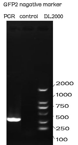
| − | + | ===PCR=== | |
| − | + | '''Methods''' | |
| − | + | The PCR is performed with Premix EX Taq by Takara. | |
| − | + | F-Prime: 5’-CGGAATTCGCGGCCGCTTCTAGAAAGAATGGAATCC-3’ | |
| + | R-Prime: 5’-GGACTAGTTTATTATTTGTATAGTTC -3’ | ||
| − | === | + | The PCR protocol is selected based on the Users Manuel. |
| + | The Electrophoresis was performed on a 1% Agarose glu. | ||
| + | |||
| + | '''Results''' | ||
| + | |||
| + | [[File:NUDT-GFP2-PCR.jpg|150px|]] | ||
| + | |||
| + | The result of the agarose electrophoresis was shown on the picture above. | ||
| + | |||
| + | ===Functional Test=== | ||
| + | |||
| + | This part is tested together with the part BBa_K1789003, in the device BBa_K1789019. | ||
| + | |||
| + | '''The result shows that this part can shine with GFP1.''' | ||
| + | |||
| + | <partinfo>BBa_K1789000 parameters</partinfo> | ||
| + | <!-- --> | ||
| + | |||
| + | |||
| + | |||
| + | |||
| + | |||
| + | ==Contribution: NUDT_CHINA 2016== | ||
| + | Author: Xinyuan Qiu | ||
| + | |||
| + | Summary: In this contribution, we further improved the characterization of BBa_ I715019 by optimizing its expression parameters. Also, we introduced another split-GFP system based on the sequence provided by BBa_ I715019 and BBa_ I715020 with a better noise-signal ratio compared to the traditional approaches. | ||
| + | |||
| + | |||
| + | ===characterization improvement=== | ||
| + | <html> | ||
| + | <p style="text-indent:22pt;"> | ||
| + | <span style="line-height:1;font-family:Perpetua;font-size:16px;">To | ||
| + | further improve the function of existing parts, we stimulate an </span><i><span style="line-height:1;font-family:Perpetua;font-size:16px;">in vivo</span></i><span style="line-height:1;font-family:Perpetua;font-size:16px;"> PPI situation, and tried to | ||
| + | optimize the culture condition for a better signal-to-noise ratio (SNR). For | ||
| + | such matter, two devices, containing split-GFP fragments and a complete or | ||
| + | spited zinc finger protein, were built under control of a lac operon controlled | ||
| + | T7 promoter. The complete zinc finger protein was to stimulate a PPI positive | ||
| + | situation, while the split one was to stimulate a PPI negative situation | ||
| + | . Fluorescence signal was detected by a microplate reader after an | ||
| + | overnight culture under various conditions.</span><span style="line-height:1;font-family:Perpetua;font-size:16px;"> </span><span style="line-height:1;font-family:Perpetua;font-size:16px;">Relative fluorescence intensity was then calculated with normalization | ||
| + | of OD</span><sub><span style="line-height:1;font-family:Perpetua;font-size:16px;">600</span></sub><span style="line-height:1;font-family:Perpetua;font-size:16px;"> value. The relative fluorescence intensity of each control | ||
| + | group was set arbitrarily at 1.0 (data not shown), and the levels of the other | ||
| + | groups were adjusted correspondingly. Results shown a better SNR under 20</span><span style="line-height:1;font-family:Perpetua;font-size:16px;">℃</span><span style="line-height:1;font-family:Perpetua;font-size:16px;"> and 0.5mM IPTG induction (Figure 3). Thus indicating that better performance | ||
| + | of such system could be expected under lower culturing temperature.</span> | ||
| + | </p> | ||
| + | <p style="text-indent:22pt;"> | ||
| + | <span style="line-height:1;font-family:Perpetua;font-size:16px;"> </span> | ||
| + | </p> | ||
| + | |||
| + | </br> | ||
| + | </html> | ||
| + | [[File:T--NUDT CHINA--introfig3.jpg|700px|center]] | ||
| + | <html> | ||
| + | </br> | ||
| + | <p> | ||
| + | <b><span style="line-height:1;font-family:Perpetua;font-size:16px;">Figure 1. Evaluation of the split-GFP system under different expression condition. </span></b> | ||
| + | </p> | ||
| + | <p> | ||
| + | <span style="line-height:1;font-family:Perpetua;font-size:16px;">Relative fluorescence intensity was calculated with normalization of the OD600 value. The relative fluorescence intensity of each control group was set arbitrarily at 1.0 (data not shown), and the levels of the other groups were adjusted correspondingly. Green fluorescence was measured under 488nm of excitation and 538nm of emission. This experiment was run in three parallel reactions, and the data represent results obtained from at least three independent experiments. *p<0.05, **p<0.01.</span> | ||
| + | </p> | ||
| + | </br> | ||
| + | |||
| + | </html> | ||
| + | |||
| + | ===Functional extension=== | ||
| + | |||
| + | <html> | ||
| + | <p style="text-indent:6.5pt;"> | ||
| + | <span style="line-height:1;font-family:Perpetua;font-size:16px;">To | ||
| + | further improve the function of split-GFP system, another method of splitting | ||
| + | GFP was introduced and tested in our project. Instead of a traditional two-part | ||
| + | split, we split the GFP protein into three fragments namely GFP10 (residues | ||
| + | 194-212), GFP11 (residues 213-233) and GFP 1-9 (residues 1-193)<sup>14</sup>. Due to their short | ||
| + | length, two small fragments can be easily fused onto proteins with less | ||
| + | affection on their folding </span><span style="line-height:1;font-family:Perpetua;font-size:16px;">(figure 2A)</span><span style="line-height:1;font-family:Perpetua;font-size:16px;">. </span> | ||
| + | </p> | ||
| + | <p> | ||
| + | <span style="line-height:1;font-family:Perpetua;font-size:16px;"> </span> | ||
| + | </p> | ||
| + | |||
| + | |||
| + | |||
| + | </br> | ||
| + | </html> | ||
| + | [[File:T--NUDT CHINA--introfig4.jpg|900px|center]] | ||
| + | <html> | ||
| + | </br> | ||
| + | <p> | ||
| + | <b><span style="line-height:1;font-family:Perpetua;font-size:16px;">Figure 2 Evaluation of two different split-GFP systems.</span></b> | ||
| + | </p> | ||
| + | <p> | ||
| + | <span style="line-height:1;font-family:Perpetua;font-size:16px;">Relative fluorescence intensity was calculated with normalization of the OD600 value. For Relative FI ratios, relative fluorescence intensity of each control group was set arbitrarily at 1.0, and the levels of the other groups were adjusted correspondingly. This experiment was run in three parallel reactions, and the data represent results obtained from at least three independent experiments. *p<0.05, **p<0.01.</span> | ||
| + | </p> | ||
| + | </br> | ||
| + | |||
| + | |||
| + | |||
| + | <p style="text-indent:22pt;"> | ||
| + | <span style="line-height:1;font-family:Perpetua;font-size:16px;"> </span> | ||
| + | </p> | ||
| + | <p style="text-indent:22pt;"> | ||
| + | <span style="line-height:1;font-family:Perpetua;font-size:16px;">Comparing | ||
| + | with previous split-GFP system, higher SNR was reached under the same | ||
| + | expression condition, while the total signal intensity suffered tolerable | ||
| + | decrease (Figure 2B).</span> | ||
| + | </p> | ||
| + | |||
| + | </html> | ||
| + | |||
| + | |||
| + | |||
| + | |||
| + | |||
| + | |||
| + | |||
| + | |||
| + | |||
| + | |||
| + | |||
| + | |||
| + | |||
| + | ==References== | ||
[ 1] Drewes G, Bouwmeester T.Global approaches to protein – protein interaction[ J ]. Curr Opin Cell Biol, 2003, 15(2): 199-205. | [ 1] Drewes G, Bouwmeester T.Global approaches to protein – protein interaction[ J ]. Curr Opin Cell Biol, 2003, 15(2): 199-205. | ||
Latest revision as of 18:05, 19 October 2016
GFP2
In order to check the functions of our system that whether two frames standing near enough so that enzymes could react easily, we divided the sequence of GFP into two parts called N-fragment and C-fragment. This is the latter. This works like this way: only when supplied with the proper distance for tow enzymes will it produce light output.
Usage and Biology
Bimolecular fluorescence complementation (BiFC) means two non-fluorescent complementary fragments of the fluorescent protein can reassemble to form a fluorescent complex and restore fluorescence when they are fused to two proteins that interact with each other.
BiFC analysis has been used to study interactions among a wide range of proteins in many cell types. The study of interactions and post-translational modification of the protein makes people master the biological regulatory mechanism more. Interactions in protein are also highly valued, so there have been a number of related technologies having different characteristics and applications[1-2].
Such as GFP,after the sequence of certain sites in interchanges with the amino-terminal or carboxy-terminal sequence loop, it could still be able to fold correctly to form the structure of the chromophore and maintain fluorescence properties[3-5].
When fusing the GFP2 with our TALE2/3, we found the additional T base on the 5’ terminal of the BBa_I715020 annoying because it may cause frameshift mutation if we just use the standard Bio-brick assembly to fuse two enzymes. And that mutation may disable the whole reporter gene. In order to meet this challenge and make this part easier to use, we designed a pair of prime to remove that additional base and make this part easier to be used.
Sequence and Features
Sequence and Features
- 10COMPATIBLE WITH RFC[10]
- 12COMPATIBLE WITH RFC[12]
- 21COMPATIBLE WITH RFC[21]
- 23COMPATIBLE WITH RFC[23]
- 25COMPATIBLE WITH RFC[25]
- 1000INCOMPATIBLE WITH RFC[1000]Illegal BsaI.rc site found at 173
Experimental Validation
This part is validated through four ways: enzyme cutting, PCR, Sequence, and functional testing
PCR
Methods
The PCR is performed with Premix EX Taq by Takara.
F-Prime: 5’-CGGAATTCGCGGCCGCTTCTAGAAAGAATGGAATCC-3’
R-Prime: 5’-GGACTAGTTTATTATTTGTATAGTTC -3’
The PCR protocol is selected based on the Users Manuel. The Electrophoresis was performed on a 1% Agarose glu.
Results
The result of the agarose electrophoresis was shown on the picture above.
Functional Test
This part is tested together with the part BBa_K1789003, in the device BBa_K1789019.
The result shows that this part can shine with GFP1.
Contribution: NUDT_CHINA 2016
Author: Xinyuan Qiu
Summary: In this contribution, we further improved the characterization of BBa_ I715019 by optimizing its expression parameters. Also, we introduced another split-GFP system based on the sequence provided by BBa_ I715019 and BBa_ I715020 with a better noise-signal ratio compared to the traditional approaches.
characterization improvement
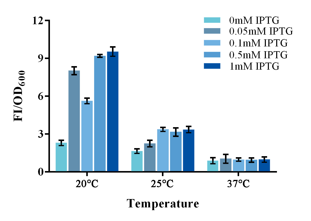
To further improve the function of existing parts, we stimulate an in vivo PPI situation, and tried to optimize the culture condition for a better signal-to-noise ratio (SNR). For such matter, two devices, containing split-GFP fragments and a complete or spited zinc finger protein, were built under control of a lac operon controlled T7 promoter. The complete zinc finger protein was to stimulate a PPI positive situation, while the split one was to stimulate a PPI negative situation . Fluorescence signal was detected by a microplate reader after an overnight culture under various conditions. Relative fluorescence intensity was then calculated with normalization of OD600 value. The relative fluorescence intensity of each control group was set arbitrarily at 1.0 (data not shown), and the levels of the other groups were adjusted correspondingly. Results shown a better SNR under 20℃ and 0.5mM IPTG induction (Figure 3). Thus indicating that better performance of such system could be expected under lower culturing temperature.
Figure 1. Evaluation of the split-GFP system under different expression condition.
Relative fluorescence intensity was calculated with normalization of the OD600 value. The relative fluorescence intensity of each control group was set arbitrarily at 1.0 (data not shown), and the levels of the other groups were adjusted correspondingly. Green fluorescence was measured under 488nm of excitation and 538nm of emission. This experiment was run in three parallel reactions, and the data represent results obtained from at least three independent experiments. *p<0.05, **p<0.01.
Functional extension
To further improve the function of split-GFP system, another method of splitting GFP was introduced and tested in our project. Instead of a traditional two-part split, we split the GFP protein into three fragments namely GFP10 (residues 194-212), GFP11 (residues 213-233) and GFP 1-9 (residues 1-193)14. Due to their short length, two small fragments can be easily fused onto proteins with less affection on their folding (figure 2A).
Figure 2 Evaluation of two different split-GFP systems.
Relative fluorescence intensity was calculated with normalization of the OD600 value. For Relative FI ratios, relative fluorescence intensity of each control group was set arbitrarily at 1.0, and the levels of the other groups were adjusted correspondingly. This experiment was run in three parallel reactions, and the data represent results obtained from at least three independent experiments. *p<0.05, **p<0.01.
Comparing with previous split-GFP system, higher SNR was reached under the same expression condition, while the total signal intensity suffered tolerable decrease (Figure 2B).
References
[ 1] Drewes G, Bouwmeester T.Global approaches to protein – protein interaction[ J ]. Curr Opin Cell Biol, 2003, 15(2): 199-205.
[ 2] Collura V, Boissy G. From protein -protein complexes to interactomics [ J ].Subcell Biochem, 2007, 43: 135-83.
[ 3] M isteli T, Spector DL. Application of the green fluorescent protein in cell biology and biotechnology[ J ].Nat Biotechnol, 1997, 15( 10): 961 - 964.
[ 4] Baird G. S, Zacharias DA, Tsien RY. Circular permutation and receptor insertion within green fluorescent proteins[ J ]. Proc Natl Acad Sci USA , 1999, 96( 20): 11241-11246.
[ 5] Ghosh I, Hamilton AD, Regan L. Antiparallel leucinezipper –directed protein reassembly: application to the green fluorescent protein[ J ]. J Am Chem Soc, 2000, 122: 5658-5659.


