Difference between revisions of "Part:BBa K771005"
(→Constitutive Aggregation) |
AleAlejandro (Talk | contribs) (→Characterization) |
||
| (14 intermediate revisions by 3 users not shown) | |||
| Line 2: | Line 2: | ||
<partinfo>BBa_K771005 short</partinfo> | <partinfo>BBa_K771005 short</partinfo> | ||
| − | Membrane anchor 5, which consists of blue light sensor VVD([https://parts.igem.org/wiki/index.php?title=Part:BBa_K771109 BBa_K771109]) , | + | Membrane anchor 5, which consists of blue light sensor VVD([https://parts.igem.org/wiki/index.php?title=Part:BBa_K771109 BBa_K771109]) ,GBD ligand([https://parts.igem.org/wiki/index.php?title=Part:BBa_K771106 BBa_K771106]) and membrane protein Lgt([https://parts.igem.org/wiki/index.php?title=Part:BBa_K771102 BBa_K771102]). Membrane Anchor 5 is proved to constitutively dimerize with Membrane Anchor 4 [[https://parts.igem.org/wiki/index.php?title=Part:BBa_K771004 BBa_K771004]] and could aggregate with Membrane Anchor 6 [[https://parts.igem.org/wiki/index.php?title=Part:BBa_K771006 BBa_K771006]] only when blue light is present. |
==Overview: Membrane Scaffold System== | ==Overview: Membrane Scaffold System== | ||
| Line 12: | Line 12: | ||
==Backgroud== | ==Backgroud== | ||
| + | [[Image:12SJTU-CA45.png|300px|center]] | ||
| − | |||
| − | + | ===Constitutive Aggregation=== | |
| − | + | Membrane Anchor 4 ([https://parts.igem.org/wiki/index.php?title=Part:BBa_K771003 BBa_K771003])and Membrane Anchor 5 (with and without VVD) constitutively aggregate through GBD domain([https://parts.igem.org/wiki/index.php?title=Part:BBa_K771105 BBa_K771105]) and GBD ligand ([https://parts.igem.org/wiki/index.php?title=Part:BBa_K771106 BBa_K771106]). | |
| − | === | + | ===Blue Light Induced Aggregation=== |
| − | + | ||
| − | + | We incorporated VVD([https://parts.igem.org/wiki/index.php?title=Part:BBa_K771109 BBa_K771109]) as a blue light sensor to control the aggregation state of Membrane Scaffold system. VVD is a photoreceptor of Neurospora crassa which can form dimer only in the presence of blue light and would disassociate as light is off. Mutant (C71V and N56K) is harder to dimerize in the dark and easier to disassociate under blue light compared with wildtype VVD. | |
| − | aggregate | + | When blue light is on, Membrane Anchor 5 and 6 aggregate, activating pathway involving enzymes 2, 3, 4, 5 and 6. |
| − | + | ||
| − | + | ||
==Characterization== | ==Characterization== | ||
| − | |||
| − | + | To testify the constitutive aggregation of Membrane Anchor 4 and Membrane Anchor 5, we conducted Fluorescence Complementation Assay. | |
| + | In fluorescence complementation assay, proteins that are postulated to interact are fused to unfolded complementary fragments of EGFP and expressed in E.coli. Interaction of these proteins will bring the fluorescent fragments within proximity, allowing the reporter protein to reform in its native three-dimensional structure and emit its fluorescent signal. EGFP was split into two halves, named 1EGFP([https://parts.igem.org/wiki/index.php?title=Part:BBa_K771113 BBa_K771113]) and 2EGFP([https://parts.igem.org/wiki/index.php?title=Part:BBa_K771114 BBa_K771114]) respectively. If there is interaction between two proteins which were fused with 1EGFP and 2EGFP, it is expected that fluorescence should be observed. Otherwise, no fluorescence could be observed. | ||
| + | ====Membrane Anchor 4 and Membrane Anchor 5 without VVD==== | ||
| + | [[Image:12SJTU_splitegfp45.png|400px|center]] | ||
| + | |||
| + | [[Image:12SJTU Standard GFP 2.jpg|400px|center]] | ||
| + | We fused split EGFP 1 and 2 to Membrane Anchor 4 and Membrane Anchor 5 without VVD to test whether Membrane Anchor 4 and Membrane Anchor 5 without VVD could constitutively aggregate. Proteins in control group are not expected to aggregate like in experimental group.We coexpressed proteins in experimental group and control group respectively in ''E.coli''. After induction for 6 hours, bacteria samples were taken for fluorescence observationThe result shows that Membrane Anchor 4 and Membrane Anchor 5 without VVD could constitutively aggregate through GBD domain and ligand. | ||
| + | |||
| + | ===Membrane Anchor 5 and Membrane Anchor 6=== | ||
| + | We also justified the blue light induced aggregation ability of Membrane Anchor 5 and 6 by recruiting [[http://2012.igem.org/Team:SJTU-BioX-Shanghai/Project/project2.1 Violacein pathway]] in our system. | ||
==Design== | ==Design== | ||
| − | [[Image:12SJTU_MA5.png| | + | [[Image:12SJTU_MA5.png|150px|center]] |
Membrane Anchor 5 could constitutively aggregate with Membrane Anchor 4 and can aggregate with Membrane Anchor 6 only when blue light is on. | Membrane Anchor 5 could constitutively aggregate with Membrane Anchor 4 and can aggregate with Membrane Anchor 6 only when blue light is on. | ||
| Line 60: | Line 65: | ||
Wang, X., X. Chen, et al. (2012). "Spatiotemporal control of gene expression by a light-switchable transgene system." Nature Methods. | Wang, X., X. Chen, et al. (2012). "Spatiotemporal control of gene expression by a light-switchable transgene system." Nature Methods. | ||
| − | |||
<!-- Add more about the biology of this part here | <!-- Add more about the biology of this part here | ||
Latest revision as of 04:51, 3 October 2012
Membrane Anchor 5:ssDsbA-VVD-LGT-GBD Ligand
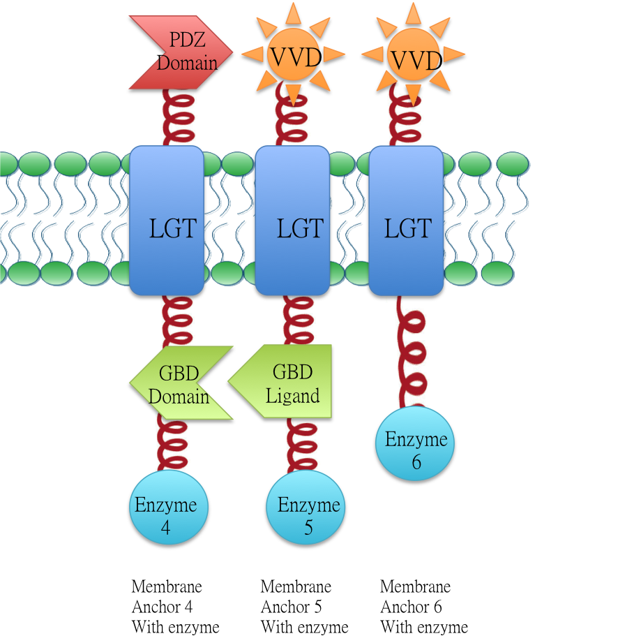
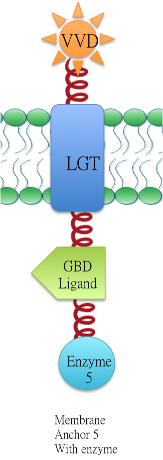
Membrane anchor 5, which consists of blue light sensor VVD(BBa_K771109) ,GBD ligand(BBa_K771106) and membrane protein Lgt(BBa_K771102). Membrane Anchor 5 is proved to constitutively dimerize with Membrane Anchor 4 [BBa_K771004] and could aggregate with Membrane Anchor 6 [BBa_K771006] only when blue light is present.
Overview: Membrane Scaffold System
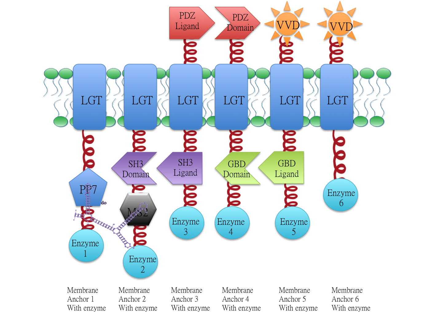
Demonstration of the whole Membrane Scaffold device. Membrane Anchor 2, 3, 4, 5 consitutively aggregate while Membrane Anchor 1 and 6 only aggregate with
constitutive cluster (Membrane Anchor 2, 3, 4, 5) when certain signal is present.
Backgroud
Constitutive Aggregation
Membrane Anchor 4 (BBa_K771003)and Membrane Anchor 5 (with and without VVD) constitutively aggregate through GBD domain(BBa_K771105) and GBD ligand (BBa_K771106).
Blue Light Induced Aggregation
We incorporated VVD(BBa_K771109) as a blue light sensor to control the aggregation state of Membrane Scaffold system. VVD is a photoreceptor of Neurospora crassa which can form dimer only in the presence of blue light and would disassociate as light is off. Mutant (C71V and N56K) is harder to dimerize in the dark and easier to disassociate under blue light compared with wildtype VVD.
When blue light is on, Membrane Anchor 5 and 6 aggregate, activating pathway involving enzymes 2, 3, 4, 5 and 6.
Characterization
To testify the constitutive aggregation of Membrane Anchor 4 and Membrane Anchor 5, we conducted Fluorescence Complementation Assay.
In fluorescence complementation assay, proteins that are postulated to interact are fused to unfolded complementary fragments of EGFP and expressed in E.coli. Interaction of these proteins will bring the fluorescent fragments within proximity, allowing the reporter protein to reform in its native three-dimensional structure and emit its fluorescent signal. EGFP was split into two halves, named 1EGFP(BBa_K771113) and 2EGFP(BBa_K771114) respectively. If there is interaction between two proteins which were fused with 1EGFP and 2EGFP, it is expected that fluorescence should be observed. Otherwise, no fluorescence could be observed.
Membrane Anchor 4 and Membrane Anchor 5 without VVD
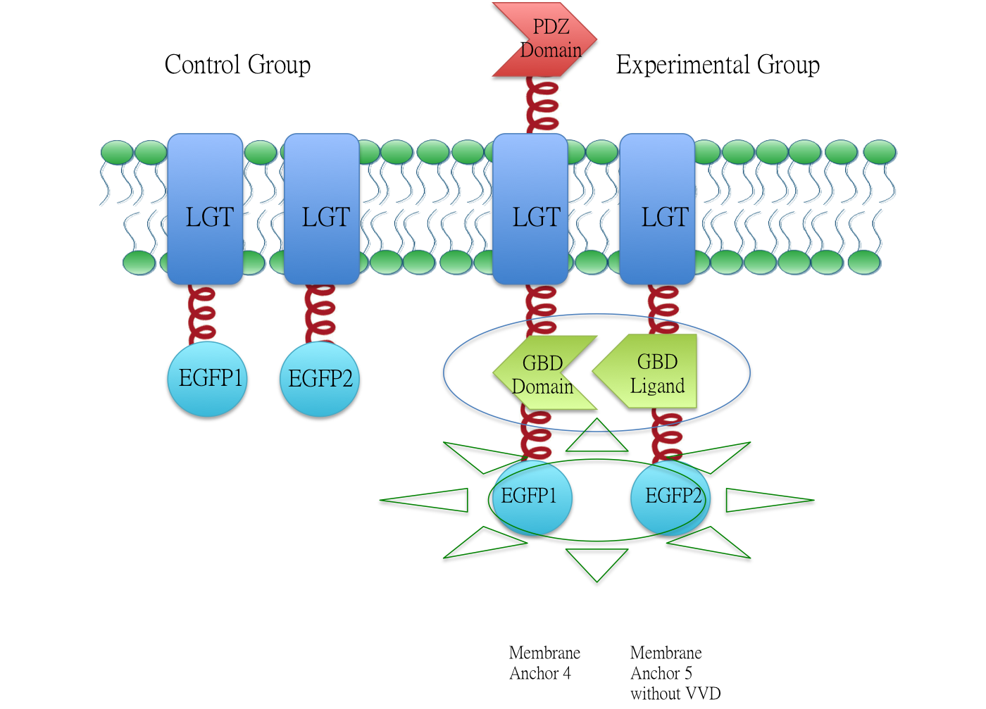
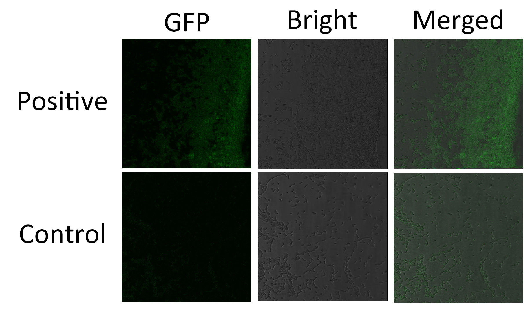
We fused split EGFP 1 and 2 to Membrane Anchor 4 and Membrane Anchor 5 without VVD to test whether Membrane Anchor 4 and Membrane Anchor 5 without VVD could constitutively aggregate. Proteins in control group are not expected to aggregate like in experimental group.We coexpressed proteins in experimental group and control group respectively in E.coli. After induction for 6 hours, bacteria samples were taken for fluorescence observationThe result shows that Membrane Anchor 4 and Membrane Anchor 5 without VVD could constitutively aggregate through GBD domain and ligand.
Membrane Anchor 5 and Membrane Anchor 6
We also justified the blue light induced aggregation ability of Membrane Anchor 5 and 6 by recruiting http://2012.igem.org/Team:SJTU-BioX-Shanghai/Project/project2.1 Violacein pathway in our system.
Design
Membrane Anchor 5 could constitutively aggregate with Membrane Anchor 4 and can aggregate with Membrane Anchor 6 only when blue light is on.
Application
We recruited Violacein biosynthetic pathway involving enzyme Vio A,B,C,D and E. Violacein biosynthetic pathway is a branched pathway which could lead to the producion of three final products.
Through blue light signal control, we successfully turned on a pathway which was otherwise almost shut off.
For more information, see BBa_K771201
Related Biobrick
<br\>Membrabe Anchor 1([BBa_K771001]) <br\>Membrabe Anchor 2([BBa_K771002]) <br\>Membrabe Anchor 3([BBa_K771003]) <br\>Membrabe Anchor 4([BBa_K771004]) <br\>Membrabe Anchor 6([BBa_K771006])
Reference
Dueber, J. E., G. C. Wu, et al. (2009). "Synthetic protein scaffolds provide modular control over metabolic flux." Nature biotechnology 27(8): 753-759.
Wang, X., X. Chen, et al. (2012). "Spatiotemporal control of gene expression by a light-switchable transgene system." Nature Methods.
Sequence and Features
- 10COMPATIBLE WITH RFC[10]
- 12COMPATIBLE WITH RFC[12]
- 21COMPATIBLE WITH RFC[21]
- 23COMPATIBLE WITH RFC[23]
- 25INCOMPATIBLE WITH RFC[25]Illegal AgeI site found at 494
Illegal AgeI site found at 1339 - 1000INCOMPATIBLE WITH RFC[1000]Illegal BsaI site found at 960

