Difference between revisions of "Part:BBa K4140008"
Ahmed Mattar (Talk | contribs) (→Experimental Characterization and verifying the construction of the improved part) |
Ahmed Mattar (Talk | contribs) (→Experimental Characterization and verifying the construction of the improved part) |
||
| Line 65: | Line 65: | ||
<br><br><br><br><br><br><br><br><br><br><br><br> | <br><br><br><br><br><br><br><br><br><br><br><br> | ||
[[File:ag1.png|right|]] | [[File:ag1.png|right|]] | ||
| − | <br><br><br><br><br><br><br><br><br><br><br><br><br><br><br><br><br><br><br><br> | + | <br><br><br><br><br><br><br><br><br><br><br><br><br><br><br><br><br><br><br><br><br><br><br><br><br> |
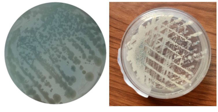
This figure shows at left (Phe / X-gal with KP-SP) +ve, shows that the WCB emitted blue color with good intensity at standard 20 mg/dL concentration of phenylalanine. and to the right (Phe / X-gal) +ve, without the tag KP-SP, shows that the WCB emitted a low level of blue color at the same concentration of phenylalanine. | This figure shows at left (Phe / X-gal with KP-SP) +ve, shows that the WCB emitted blue color with good intensity at standard 20 mg/dL concentration of phenylalanine. and to the right (Phe / X-gal) +ve, without the tag KP-SP, shows that the WCB emitted a low level of blue color at the same concentration of phenylalanine. | ||
Revision as of 08:01, 11 October 2022
lacZ alpha
Part Description
The LacZ-alpha fragment is encoded by this region and is derived from the pUC19 cloning vector. Because it is smaller than the LacZ-omega region, the LacZ-alpha fragment can be easily incorporated into a plasmid when the two non-functional LacZ gene fragments (alpha and omega) are co-expressed. X-gal (5-bromo-4-chloro-3-indoyl-d-galactopyranoside), a soluble colourless molecule that is a substrate of ß-galactosidase and generates a blue product when cleaved, is used to detect the alpha-complementation
Usage
alpha domain of ß-galactosidase (our reporter protein) which is produced in response to high levels of phenylalanine and generates a blue product when X-gal is cleaved which is a soluble colorless molecule that is a substrate of ß-galactosidase and generates a blue product when cleaved, is used to detect the alpha-complementation. We used beta-galactosidase enzyme to turn the color into blue once increased level of phenylalanine is detected via exported extracellularly via tagging by kp-sp to bin with its substrate (X-gal).
Improvements
We improved the part BBa_E0033 by :-
Improvement by KP-SP tagging
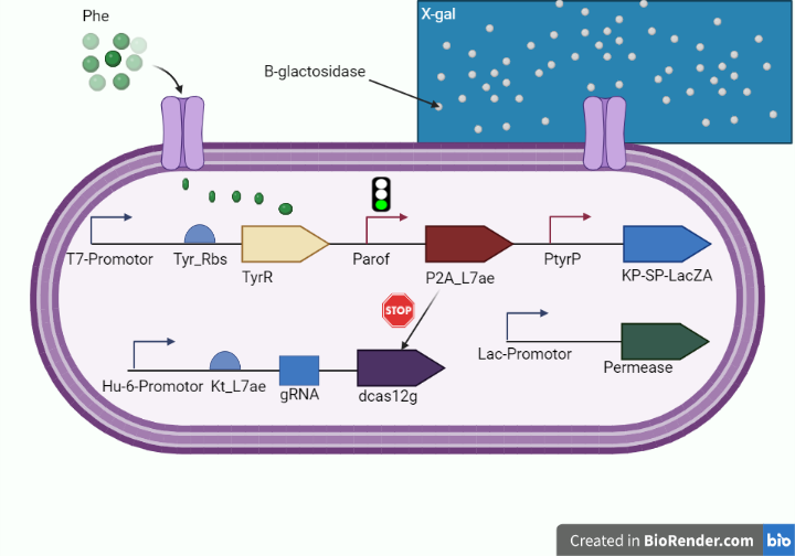
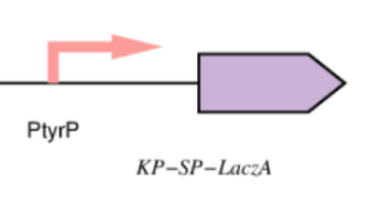
This year, we enhanced the LacZ alpha gene by incorporating a peptide signal that controls the secretion of B galactosidase extracellularly (KP-SP tagging) in an effort to improve the cleavage capacity of the X gal, heighten the intensity of the dark blue color emitted by the X gal product, and reduce the amount of time required to receive the results after performing the test on the chip. as shown in figure 1 and figure 2.
Improvement of LacZ alpha gene which encodes for the alpha domain of B-galactosidase that acts as a reporter protein in our diagnostic circuit :
Lactose converting enzyme (B-gal) beta-galactosidase that is originated from bacteria And have been studied as a reporter protein for many years ,It's a polypeptide of 1029 amino acid, and its tetramerization carries out the functional form of the enzyme.
The deletion of the tetramerization domain which is the N-terminal region spanning the first 50 residues will abolish its enzymatic activity, which could be fully restored by the alpha complementation of the intact N- terminal portion of the (Beta-gal) this alpha complementation is detected by using a beta-galactosidase colorless substrate as X gal to be cleaved into a dark blue product.
This year we employed this approach to indicate the presence of phenylalanine within our whole cell biosensor on the test line with media containing X gal substrate of our platform to diagnose phenylketonuria.
Figure 1. (shows the diagnostic whole cell-based biosensor after tagging lacZ alpha with kp-sp.)
Improvement by Directed Evolution
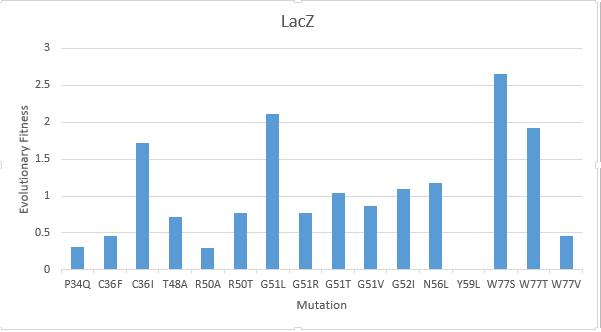
After creating a multiple sequence alignment of the protein sequence and predicting mutational landscapes, the effect of these mutations on the evolutionary fitness of the protein is tested. The prediction of the mutational landscape by saturation mutagenesis of the LacZ protein. The (W77S ) mutation, as depicted in the chart, had the greatest score when compared to other mutations. On the other hand, it's clear that the (Y59L) had the least evolutionary fitness for LacZ protein. As displayed in Figure(3)
Literature Characterization
According to the results, The ideal pH and temperature for Nf-LacZ are 40 °C and pH 6.5, respectively. The activity of the beta -galactosidase may also be impacted by metal ions. The metal ions K+, Mg2+, Ca2+, Zn2+, and Mn2+ were discovered to all be able to increase the enzymatic activity of Nf-LacZ.,and its Km was 05mmol/litre for Nf-LacZ, on the other hand the optimal temperature and PH for WspA are, respectively, 45 °C and pH 8.0, and Km value is close to to that of for WspA and this is showin in Nf-LacZ and WspA protein expression in vitro and analyzing its activity as shown in figures 4 and 5.
Characterization by mathematical modeling
We are using mathematical modeling to detect the increased level of phenylalanine (phe) in phenylketonuria patients in our diagnostic platform. It depends on a whole cell-based biosensor through a cascade of reactions to finally end by formation of β-galactosidase that turns the color into blue once bound to its substrate (X-gal) as mentioned in figure (4) and graph (1).
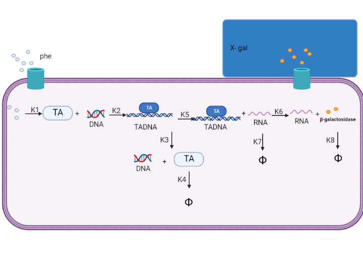
Figure (4) represents the cascade of reactions in whole cell-based biosensor model.
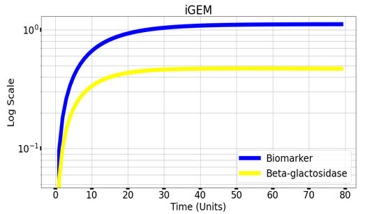
Graph(1) illustrates a direct relation between biomarker and beta-galactosidase ,so as the biomarker increases, the released amount of beta-galactosidase increases till it reaches constant value after about 30 time units. Therefore, the maximum amount of the biomarker releases the maximum amount of beta-galactosidase.
Experimental Characterization and verifying the construction of the improved part
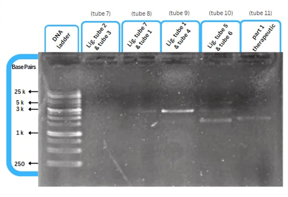
This figure shows an experimental characterization of this part as it's validated through gel electrophoresis as it is in lane 3. The running part (ordered from IDT) included KP-SP and lacZ alpha.
This figure shows at left (Phe / X-gal with KP-SP) +ve, shows that the WCB emitted blue color with good intensity at standard 20 mg/dL concentration of phenylalanine. and to the right (Phe / X-gal) +ve, without the tag KP-SP, shows that the WCB emitted a low level of blue color at the same concentration of phenylalanine.
References
1.Liu, W., Cui, L., Xu, H., Zhu, Z. & Gao, X. Flexibility-rigidity coordination of the dense exopolysaccharide matrix in terrestrial cyanobacteria acclimated to periodic desiccation. Appl. Environ. Microbiol. 83, e01619–17 (2017). 2.Wright, D. J. et al. UV irradiation and desiccation modulate the three-dimensional extracellular matrix of Nostoc commune (Cyanobacteria). J. Biol. Chem. 280, 40271–40281 (2005).
Sequence and Features
- 10COMPATIBLE WITH RFC[10]
- 12COMPATIBLE WITH RFC[12]
- 21INCOMPATIBLE WITH RFC[21]Illegal BamHI site found at 93
Illegal XhoI site found at 144 - 23COMPATIBLE WITH RFC[23]
- 25COMPATIBLE WITH RFC[25]
- 1000COMPATIBLE WITH RFC[1000]

