
Difference between revisions of "Part:BBa K1800000:Experience"
| Line 4: | Line 4: | ||
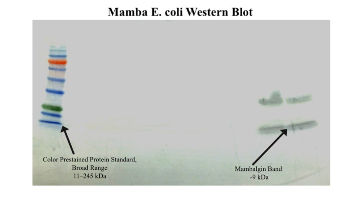
| − | The 2015 GSU iGEM team was able to successfully express and characterize the mambalgin protein in E. coli. After inducing with IPTG overnight, the cell cultures were centrifuged and disrupted with a French press. Mambalgin was isolated using affinity chromatography, utilizing the 6x His tag in the construct. A | + | The 2015 GSU iGEM team was able to successfully express and characterize the mambalgin protein in E. coli. After inducing with IPTG overnight, the cell cultures were centrifuged and disrupted with a French press. Mambalgin was isolated using affinity chromatography, utilizing the 6x His tag in the construct. A coomassie stain was performed to visualize the proteins. A band indicating a protein of ~9 kda – the size of the mambalgin for E. coli construct – was seen in the eluate fraction which is indicative of our desired protein. However, this band may also contain any native proteins that migrate below 11 kD. An anti-myc Western was performed to confirm the presence of mambalgin and a signal was observed at the expected size. An additional band at ~35 kd was detected however. We believe that this may be due to aggregates formed by non-specific disulfide bond formation. |
<br> | <br> | ||
https://static.igem.org/mediawiki/parts/d/d9/Gsuigemgel.png | https://static.igem.org/mediawiki/parts/d/d9/Gsuigemgel.png | ||
Revision as of 23:31, 26 September 2015
This experience page is provided so that any user may enter their experience using this part.
Please enter
how you used this part and how it worked out.
The 2015 GSU iGEM team was able to successfully express and characterize the mambalgin protein in E. coli. After inducing with IPTG overnight, the cell cultures were centrifuged and disrupted with a French press. Mambalgin was isolated using affinity chromatography, utilizing the 6x His tag in the construct. A coomassie stain was performed to visualize the proteins. A band indicating a protein of ~9 kda – the size of the mambalgin for E. coli construct – was seen in the eluate fraction which is indicative of our desired protein. However, this band may also contain any native proteins that migrate below 11 kD. An anti-myc Western was performed to confirm the presence of mambalgin and a signal was observed at the expected size. An additional band at ~35 kd was detected however. We believe that this may be due to aggregates formed by non-specific disulfide bond formation.

Applications of BBa_K1800000
User Reviews
UNIQaba90d120a51a9f2-partinfo-00000000-QINU UNIQaba90d120a51a9f2-partinfo-00000001-QINU