
Difference between revisions of "Part:BBa K1323016"
| (5 intermediate revisions by the same user not shown) | |||
| Line 2: | Line 2: | ||
<partinfo>BBa_K1323016 short</partinfo> | <partinfo>BBa_K1323016 short</partinfo> | ||
| − | The Translation Initiation Region ribosome binding site (TIR rbs) was isolated from T7 bacteriophage (Fey, 2014). It is used for optimal expression in | + | The Translation Initiation Region ribosome binding site (TIR rbs) was isolated from T7 bacteriophage (Fey, 2014). It is used for optimal expression in ''S.epidermidis'' when a native RBS is too poor to achieve desired expression levels. It has a Shine-Dalgarno consensus sequence (AGGAGG). The start codon and the phage T7 enhancer were spaced to optimally minimize secondary structure formation (Cheng et al., 1992). The TIR RBS leads to greater expression levels compared to ''sodA'' RBS [https://parts.igem.org/Part:BBa_K1323022 BBa_K1323022] even though they have the same consensus sequence (Fey, 2014). |
| − | + | (A)[[File:DsRED.png]] (B)[[File:DsRED!.tif]] | |
| − | + | DsRed fluorescence was analyzed in both E. coli and ''S. epidermidis''. The DsRed cassette was incorporated into a pUC57 plasmid containing an erythromycin resistance gene ([https://parts.igem.org/Part:BBa_K1323011 BBa_K1323011]) and Staphylococcal oriV ([https://parts.igem.org/Part:BBa_K1323018 BBa_K1323018]). This plasmid was transformed into the strains using heat shock for E. coli and electroporation for ''S. epidermidis''. Pellets from 50 ml overnight cultures were collected into 1.5 ml eppendorf tubes. To the naked eye (A) E. coli has a pink color (left), whereas in ''S. epidermidis'' (middle), a slight pink colour could only be noticed if compared to ''S. epidermidis'' without the construct (right). When seen under a RFP filter (B) a high degree of red fluorescence was observed in E. coli (left) and a lower degree of red fluorescence was seen in ''S. epidermidis'' (middle). The ''S. epidermidis'' negative control (right) showed no observable fluorescence. | |
| − | |||
<!-- Add more about the biology of this part here | <!-- Add more about the biology of this part here | ||
| Line 17: | Line 16: | ||
<span class='h3bb'>Sequence and Features</span> | <span class='h3bb'>Sequence and Features</span> | ||
<partinfo>BBa_K1323016 SequenceAndFeatures</partinfo> | <partinfo>BBa_K1323016 SequenceAndFeatures</partinfo> | ||
| + | |||
| + | |||
| + | == References == | ||
| + | |||
| + | Cheng, X., Patterson, T. A., (1992) Construction and use of lambda PL promoter vectors for direct cloning and high level expression of PCR amplified DNA coding sequences. Nucleic Acids Res 20, 4591–4598. | ||
| + | |||
| + | Fey, P. D. (2014). Staphylococcus epidermidis: methods and protocols. New York: Springer Science + Business Media, LLC. | ||
Latest revision as of 16:23, 2 November 2014
Translational initiation region (RBS) for Staphylococcus aureus
The Translation Initiation Region ribosome binding site (TIR rbs) was isolated from T7 bacteriophage (Fey, 2014). It is used for optimal expression in S.epidermidis when a native RBS is too poor to achieve desired expression levels. It has a Shine-Dalgarno consensus sequence (AGGAGG). The start codon and the phage T7 enhancer were spaced to optimally minimize secondary structure formation (Cheng et al., 1992). The TIR RBS leads to greater expression levels compared to sodA RBS BBa_K1323022 even though they have the same consensus sequence (Fey, 2014).
(A) (B)
(B)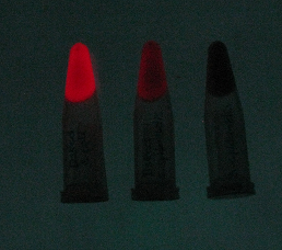
DsRed fluorescence was analyzed in both E. coli and S. epidermidis. The DsRed cassette was incorporated into a pUC57 plasmid containing an erythromycin resistance gene (BBa_K1323011) and Staphylococcal oriV (BBa_K1323018). This plasmid was transformed into the strains using heat shock for E. coli and electroporation for S. epidermidis. Pellets from 50 ml overnight cultures were collected into 1.5 ml eppendorf tubes. To the naked eye (A) E. coli has a pink color (left), whereas in S. epidermidis (middle), a slight pink colour could only be noticed if compared to S. epidermidis without the construct (right). When seen under a RFP filter (B) a high degree of red fluorescence was observed in E. coli (left) and a lower degree of red fluorescence was seen in S. epidermidis (middle). The S. epidermidis negative control (right) showed no observable fluorescence.
Sequence and Features
- 10COMPATIBLE WITH RFC[10]
- 12COMPATIBLE WITH RFC[12]
- 21COMPATIBLE WITH RFC[21]
- 23COMPATIBLE WITH RFC[23]
- 25COMPATIBLE WITH RFC[25]
- 1000COMPATIBLE WITH RFC[1000]
References
Cheng, X., Patterson, T. A., (1992) Construction and use of lambda PL promoter vectors for direct cloning and high level expression of PCR amplified DNA coding sequences. Nucleic Acids Res 20, 4591–4598.
Fey, P. D. (2014). Staphylococcus epidermidis: methods and protocols. New York: Springer Science + Business Media, LLC.