
Difference between revisions of "Part:BBa K1323009"
| (9 intermediate revisions by 2 users not shown) | |||
| Line 2: | Line 2: | ||
<partinfo>BBa_K1323009 short</partinfo> | <partinfo>BBa_K1323009 short</partinfo> | ||
| − | This is the coding sequence of DsRed_T3 (DNT), a highly fluorescent mutant version of a red fluorescent protein originally found in Discosoma (corals) (Dunn et. al., 2006). It was codon optimized for Staphylococcus aureus by Bose et. al. (2013) and we had this part synthesized by Bio Basic Canada Inc. | + | This is the coding sequence of DsRed_T3 (DNT), a highly fluorescent mutant version of a red fluorescent protein originally found in Discosoma (corals) (Dunn et. al., 2006). It was codon optimized for ''Staphylococcus aureus'' by Bose et. al. (2013) and we had this part synthesized by Bio Basic Canada Inc. |
| − | + | (A)[[File:DsRED.png]] (B)[[File:DsRED!.tif]] | |
| − | + | DsRed fluorescence was analyzed in both E. coli and ''S. epidermidis''. The DsRed cassette ([https://parts.igem.org/Part:BBa_K1323015 BBa_K1323015]) was incorporated into a pUC57 plasmid containing an erythromycin resistance gene ([https://parts.igem.org/Part:BBa_K1323011 BBa_K1323011]) and Staphylococcal oriV ([https://parts.igem.org/Part:BBa_K1323018 BBa_K1323018]). This plasmid was transformed into the strains using heat shock for E. coli and electroporation for ''S. epidermidis''. Pellets from 50 ml overnight cultures were collected into 1.5 ml eppendorf tubes. To the naked eye (A) E. coli has a pink color (left), whereas in ''S. epidermidis'' (middle), a slight pink colour could only be noticed if compared to ''S. epidermidis'' without the construct (right). When seen under a RFP filter (B) a high degree of red fluorescence was observed in E. coli (left) and a lower degree of red fluorescence was seen in ''S. epidermidis'' (middle). The ''S. epidermidis'' negative control (right) showed no observable fluorescence. | |
| − | + | ||
| − | + | ||
| − | + | ||
| − | + | ||
| − | + | ||
| − | + | ||
<!-- --> | <!-- --> | ||
<span class='h3bb'>Sequence and Features</span> | <span class='h3bb'>Sequence and Features</span> | ||
<partinfo>BBa_K1323009 SequenceAndFeatures</partinfo> | <partinfo>BBa_K1323009 SequenceAndFeatures</partinfo> | ||
| + | |||
| + | |||
| + | |||
| + | ==References== | ||
| + | |||
| + | Bose, J. L., Fey, P. D., and Bayles, K. W. (2013). Genetic Tools to Enhance the Study of Gene Function and Regulation in Staphylococcus aureus. ''Applied and Environmental Microbiology''. '''79''' (7): 2218-2224. | ||
| + | |||
| + | Dunn, A. K., Millikan, D. S., Adin, D. M., Bose, J. B., and Stabb, E. V. (2006). New rfp- and pES213-Derived Tools for Analyzing Symbiotic Vibrio fisheri Reveal Patterns of Infection and lux Expression In Situ. ''Applied and Environmental Microbiology''. '''72''' (1): 802-810. | ||
Latest revision as of 02:34, 18 October 2014
DsRed (RFP) Coding Sequence
This is the coding sequence of DsRed_T3 (DNT), a highly fluorescent mutant version of a red fluorescent protein originally found in Discosoma (corals) (Dunn et. al., 2006). It was codon optimized for Staphylococcus aureus by Bose et. al. (2013) and we had this part synthesized by Bio Basic Canada Inc.
(A) (B)
(B)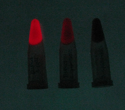
DsRed fluorescence was analyzed in both E. coli and S. epidermidis. The DsRed cassette (BBa_K1323015) was incorporated into a pUC57 plasmid containing an erythromycin resistance gene (BBa_K1323011) and Staphylococcal oriV (BBa_K1323018). This plasmid was transformed into the strains using heat shock for E. coli and electroporation for S. epidermidis. Pellets from 50 ml overnight cultures were collected into 1.5 ml eppendorf tubes. To the naked eye (A) E. coli has a pink color (left), whereas in S. epidermidis (middle), a slight pink colour could only be noticed if compared to S. epidermidis without the construct (right). When seen under a RFP filter (B) a high degree of red fluorescence was observed in E. coli (left) and a lower degree of red fluorescence was seen in S. epidermidis (middle). The S. epidermidis negative control (right) showed no observable fluorescence.
Sequence and Features
- 10COMPATIBLE WITH RFC[10]
- 12COMPATIBLE WITH RFC[12]
- 21COMPATIBLE WITH RFC[21]
- 23COMPATIBLE WITH RFC[23]
- 25COMPATIBLE WITH RFC[25]
- 1000COMPATIBLE WITH RFC[1000]
References
Bose, J. L., Fey, P. D., and Bayles, K. W. (2013). Genetic Tools to Enhance the Study of Gene Function and Regulation in Staphylococcus aureus. Applied and Environmental Microbiology. 79 (7): 2218-2224.
Dunn, A. K., Millikan, D. S., Adin, D. M., Bose, J. B., and Stabb, E. V. (2006). New rfp- and pES213-Derived Tools for Analyzing Symbiotic Vibrio fisheri Reveal Patterns of Infection and lux Expression In Situ. Applied and Environmental Microbiology. 72 (1): 802-810.