Difference between revisions of "Part:BBa K1420005"
| Line 18: | Line 18: | ||
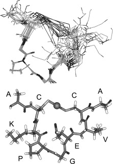
Fig 2. Structure of MerT bound to Hg(II). Top Panel: Images of the 20 lowest energy states that have been superimposed upon each other. Bottom Panel: Lowest energy structure of MerT in which the sphere represents the mercury. Reference: ''E. Rossy et al''. FEBS Lett. (2004) 575, 86–90. | Fig 2. Structure of MerT bound to Hg(II). Top Panel: Images of the 20 lowest energy states that have been superimposed upon each other. Bottom Panel: Lowest energy structure of MerT in which the sphere represents the mercury. Reference: ''E. Rossy et al''. FEBS Lett. (2004) 575, 86–90. | ||
| + | |||
<b> Mechanism </b> | <b> Mechanism </b> | ||
| Line 23: | Line 24: | ||
The transport mechanism of ionic mercury from the bacterial cell periplasm to cytosplasm is not completely understood. Hg(II) is transferred from the periplasmic cysteine pair on the first transmembrane helix to the cytoplasmic loop cysteine, where it is finally transferred to a cysteine pair at the N-terminus of the protein. The Hg(II) may go directly to MerA from the N-terminus of MerT or transfer to cytoplasmic low-molecular mass thiols. | The transport mechanism of ionic mercury from the bacterial cell periplasm to cytosplasm is not completely understood. Hg(II) is transferred from the periplasmic cysteine pair on the first transmembrane helix to the cytoplasmic loop cysteine, where it is finally transferred to a cysteine pair at the N-terminus of the protein. The Hg(II) may go directly to MerA from the N-terminus of MerT or transfer to cytoplasmic low-molecular mass thiols. | ||
| − | |||
| − | |||
| + | <b> Experimental Results </b> | ||
| + | |||
| + | [[File:MNiGEM_ZOI_MerP.jpg|center|400px|x]] | ||
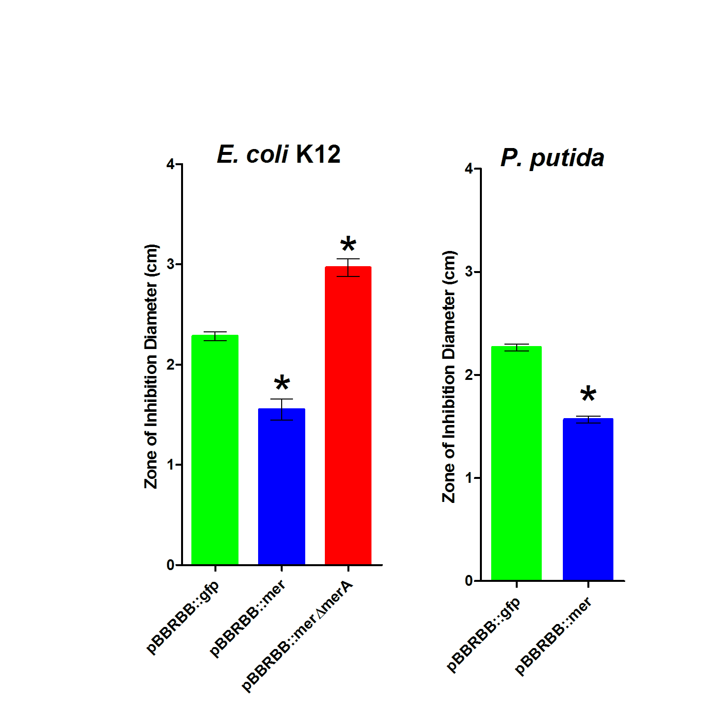
| + | Figure 3. Zone of Inhibition Plate Assasys. Strains were tested in triplicate and included ''E. coli'' K12 pBBRBB::''mer'', ''E. coli'' K12 pBBRBB::''gfp'', ''E. coli'' K12 pBBRBB::''merΔmerA'' (mer operon with ''merA'' deleted), ''P. putida'' pBBRBB::''mer'', and ''P. putida'' pBBRBB::''gfp''. | ||
| + | |||
| + | |||
| + | To complete zone of inhibition plate assays, 10µL of a 0.1M HgCl<sub>2</sub> solution was spotted on a filter disk in the middle of an agar plate. Bacteria that are resistant to mercury are able to grow closer to the disc and hence have smaller zones of inhibition. On the other hand, bacteria that are sensitive to mercury are not able to grow near the filter disc resulting in a larger zone of inhibition and hence a larger diameter. As shown in Figure 3, for ''E. coli'', the strain harboring the ''mer'' operon (pBBRBB::''mer'') had the smallest zones of inhibition indicating enhanced resistance to Hg(II) as compared to the negative control (pBBRBB::''gfp''). In ''E. coli'' containing the ''mer'' operon with ''merA'' deleted (pBBRBB:''merΔmerA''), the zone of inhibition was larger than the negative control. This result is due to the fact that MerT and MerP facilitate mercury transport into cells, but without MerA, cells are unable to detoxify/reduce Hg(II) to Hg(0) leading to increased sensitivity. Increased sensitivity to Hg(II) with ''merT'' and ''merP'' in the absence of ''merA'' indicate that MerT and MerP are functional as Hg(II) transporters. | ||
| + | |||
| + | |||
| + | <b>Sequence and Features</b> | ||
<!-- --> | <!-- --> | ||
<partinfo>BBa_K1420005 SequenceAndFeatures</partinfo> | <partinfo>BBa_K1420005 SequenceAndFeatures</partinfo> | ||
| − | + | ||
<!-- --> | <!-- --> | ||
Revision as of 08:36, 17 October 2014
merT, mercuric transport protein
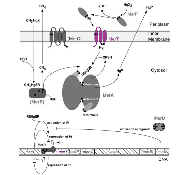
Transmembrane mercuric binding gene merT (0.3Kb) encodes MerT, which transports Hg(II) species from the periplasm through the membrane. MerT and gene merT are highlighted in purple in Figure 1. The lower part of the Figure 1 shows the arrangement of mer genes in the operon, and merT is located upstream of merP, another gene that encodes a mercury transport protein. The gene merT is found in the Serratia marcescens plasmid pDU1358 downstream of the bidirectional promoter of merR, and it is 351 base pairs long.
Figure 1. Model of transmembrane mercuric binding protein MerT. The symbol • indicates a cysteine residue. RSH indicates cytosolic thiol redox buffers such as glutathione. Figure 1 shows the interactions of MerT, in purple, with mercury compounds and other gene products of mer operon. (This figure is adapted from "Bacterial mercury resistance from atoms to ecosystems". Reference: T. Barkay et al. FEMS Microbiology Reviews 27 (2003) 355-384.)
Protein
MerT is a 116-residue (12.4 kDa) transmembrane protein. The exact structure is not known, but the protein is predicted to have three transmembrane helices with a cysteine pair accessible from the periplasmic side and another pair on the cytoplasmic side.
Fig 2. Structure of MerT bound to Hg(II). Top Panel: Images of the 20 lowest energy states that have been superimposed upon each other. Bottom Panel: Lowest energy structure of MerT in which the sphere represents the mercury. Reference: E. Rossy et al. FEBS Lett. (2004) 575, 86–90.
Mechanism
The transport mechanism of ionic mercury from the bacterial cell periplasm to cytosplasm is not completely understood. Hg(II) is transferred from the periplasmic cysteine pair on the first transmembrane helix to the cytoplasmic loop cysteine, where it is finally transferred to a cysteine pair at the N-terminus of the protein. The Hg(II) may go directly to MerA from the N-terminus of MerT or transfer to cytoplasmic low-molecular mass thiols.
Experimental Results
Figure 3. Zone of Inhibition Plate Assasys. Strains were tested in triplicate and included E. coli K12 pBBRBB::mer, E. coli K12 pBBRBB::gfp, E. coli K12 pBBRBB::merΔmerA (mer operon with merA deleted), P. putida pBBRBB::mer, and P. putida pBBRBB::gfp.
To complete zone of inhibition plate assays, 10µL of a 0.1M HgCl2 solution was spotted on a filter disk in the middle of an agar plate. Bacteria that are resistant to mercury are able to grow closer to the disc and hence have smaller zones of inhibition. On the other hand, bacteria that are sensitive to mercury are not able to grow near the filter disc resulting in a larger zone of inhibition and hence a larger diameter. As shown in Figure 3, for E. coli, the strain harboring the mer operon (pBBRBB::mer) had the smallest zones of inhibition indicating enhanced resistance to Hg(II) as compared to the negative control (pBBRBB::gfp). In E. coli containing the mer operon with merA deleted (pBBRBB:merΔmerA), the zone of inhibition was larger than the negative control. This result is due to the fact that MerT and MerP facilitate mercury transport into cells, but without MerA, cells are unable to detoxify/reduce Hg(II) to Hg(0) leading to increased sensitivity. Increased sensitivity to Hg(II) with merT and merP in the absence of merA indicate that MerT and MerP are functional as Hg(II) transporters.
Sequence and Features
- 10COMPATIBLE WITH RFC[10]
- 12INCOMPATIBLE WITH RFC[12]Illegal NheI site found at 45
- 21COMPATIBLE WITH RFC[21]
- 23COMPATIBLE WITH RFC[23]
- 25COMPATIBLE WITH RFC[25]
- 1000INCOMPATIBLE WITH RFC[1000]Illegal SapI site found at 30


