Featured Parts:Fluorescent proteins
Fluorescent proteins (FP) are convenient ways to visualize or quantify the output of a device or part. Many different FP have been cloned for use in the Registry.
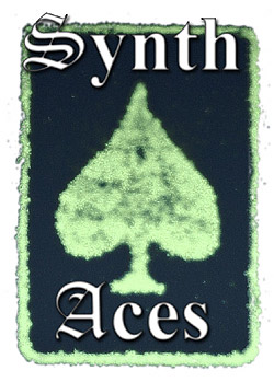
The first FP to be cloned, was green fluorescent protein (GFP), shown in the photo on the left, which as its name implies gives its biological origin (a jellyfish) a green glow. From this starting place, several investigators generated mutations that altered the spectral properties of the FP. Now we have enhanced yellow fluorescent protein (EYFP), cyan fluorescent protein (CFP) and GFP.
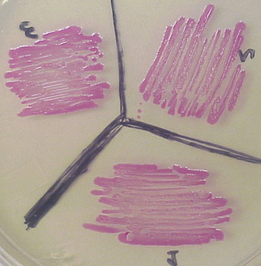
Red fluorescent protein (RFP), shown on the right, was isolated from a very different species (a coral) and its sequence is very different from the GFP family of FP. Several derivatives of RFP have been generated, most notably mOrange and mCherry.
One more technique you should know about is the use of degradation tags; LVA and AAV. These short amino acid tags (~11 amino acids) are added to the carboxyl terminus to increase the rate of protein degradation and thus facilitate a shorter half-life of the tagged protein. These degradation tags work in prokaryotes and allow you to “turn off” an FP in favor of another reporter state. This was used in the classic repressilator of Elowitz and Leibler (2000).
If you want to compare different FPs, a series of comparable [http://2006.igem.org/Davidson_Part_Nomenclature plasmids have been generated] to permit standardization of FPs. This work began with part BBa_I6060 which had a reputation as an unreliable FP. As you can read from the I6060 page, some aspects of FP are still not well documented.
More Information/References
[http://www.nature.com/nbt/journal/v22/n12/full/nbt1204-1524.html Patterson (2004). "A new harvest of fluorescent proteins" (review), Nature Biotechnology 22, 1524 - 1525.]
[http://www.bio.davidson.edu/courses/Molbio/restricted/02GFPseq/GFPseq.html Prashera, et al. (1992). "Primary structure of the Aequorea victoria green-fluorescent protein," Gene, 111, 229-233.]
[http://www.bio.davidson.edu/courses/genomics/method/GFP.html Web page about GFP with interactive Jmol image.]
[http://www.bio.davidson.edu/courses/Molbio/restricted/02GFPwow/GFPwowpg1.html Chalfie, et al. (1994). "Green Fluorescent Protein as a Marker for Gene Expression," Science 263, 802-805.]
[http://www.nature.com/cgi-taf/DynaPage.taf?file=/nature/journal/v403/n6767/abs/403335a0_fs.html&dynoptions=doi1043774228 Elowitz and Leibler (2000). "A synthetic oscillatory network of transcriptional regulators," Nature, 403: 335 - 338.]
[http://www.pubmedcentral.gov/articlerender.fcgi?tool=pubmed&pubmedid=11050229 Baird, et al. (2000). "Biochemistry, mutagenesis, and oligomerization of DsRed, a red fluorescent protein from coral," Proc. Natl. Acad. Sci. USA 97(22): 11984–11989.]


