File list
This special page shows all uploaded files.
| Date | Name | Thumbnail | Size | Description | Versions |
|---|---|---|---|---|---|
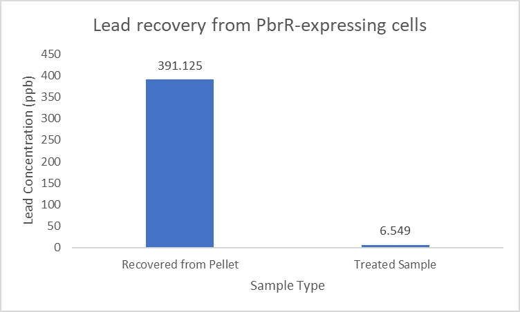
| 15:14, 12 October 2022 | IITD PbrR Recovery.png (file) |  |
9 KB | 1 | |
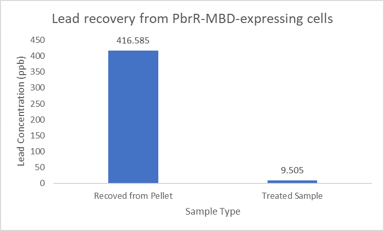
| 15:14, 12 October 2022 | IITD PbrR MBD Recovery.png (file) |  |
9 KB | 1 | |
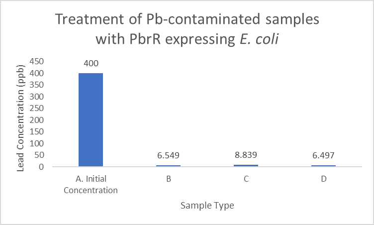
| 15:13, 12 October 2022 | IITD PbrR Treatment.png (file) |  |
11 KB | 1 | |
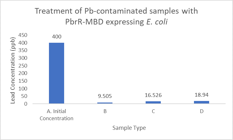
| 15:12, 12 October 2022 | IITD PbrR MBD Treatment.png (file) |  |
10 KB | Improves lead removal | 1 |
| 21:51, 9 October 2022 | IITD PbrR691 Adsorption Capacity.png (file) |  |
33 KB | Relative adsorption capacities for different mixed metal concentrations for PbrR, PbrR691, and PbrD (Jia et al., 2020) | 1 |
| 21:41, 9 October 2022 | IITD PbrR691 3D Structure.png (file) |  |
47 KB | Protein structure of PbrR691 | 1 |
| 21:31, 9 October 2022 | IITD PbrR MBD Display.png (file) |  |
292 KB | Surface display of PbrR and PbBD fusions to LOA in E. coli BL21(DE3). a Bright field and corresponding fluorescent micrographs of surface-engineered cells. Immunofluorescence labelling of E. coli cells using anti-His tag antibody (primary antibody) and... | 1 |
| 21:21, 9 October 2022 | IITD PbrR MBD Structure.png (file) |  |
265 KB | A model of PbrR protomer generated using Swiss-Model workspace (https://swissmodel.expasy.org/workspace/). The ribbon diagram shows the Pb2+ binding domain in color and the other parts in gray. Residues Cys79, Cys114, and Cys123 involved in metal bindi... | 1 |
