
File:TU Eindhoven design ompxstructure.png
No higher resolution available.
TU_Eindhoven_design_ompxstructure.png (500 × 271 pixels, file size: 82 KB, MIME type: image/png)
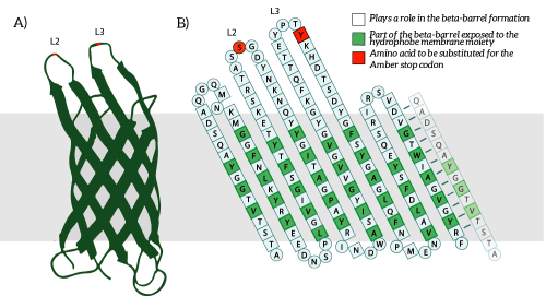
A) The OmpX protein structure has been elucidated through NMR and X-ray crystallography, B) The square residues are important for the secondary structure of OmpX. To keep the structure intact, we introduce an amber stop codon in one of the protruding loops. Figure 5B is adapted from [1]. [1] J. Vogt and G. E. Schulz, “The structure of the outer membrane protein OmpX from Escherichia coli reveals possible mechanisms of virulence.,” Structure, vol. 7, no. 10, pp. 1301–9, Oct. 1999.
File history
Click on a date/time to view the file as it appeared at that time.
| Date/Time | Thumbnail | Dimensions | User | Comment | |
|---|---|---|---|---|---|
| current | 14:10, 9 September 2015 | | 500 × 271 (82 KB) | S136969 (Talk | contribs) | A) The OmpX protein structure has been elucidated through NMR and X-ray crystallography, B) The square residues are important for the secondary structure of OmpX. To keep the structure intact, we introduce an amber stop codon in one of the protruding loop |
- You cannot overwrite this file.
File usage
The following 6 pages link to this file: